. 2023 Aug 23:14:1230050.
doi: 10.3389/fimmu.2023.1230050. eCollection 2023.

No NLRP3 inflammasome activity in kidney epithelial cells, not even when the NLRP3-A350V Muckle-Wells variant is expressed in podocytes of diabetic mice

Affiliations

No NLRP3 inflammasome activity in kidney epithelial cells, not even when the NLRP3-A350V Muckle-Wells variant is expressed in podocytes of diabetic mice

Sophie Carina Kunte et al. Front Immunol. .

Abstract

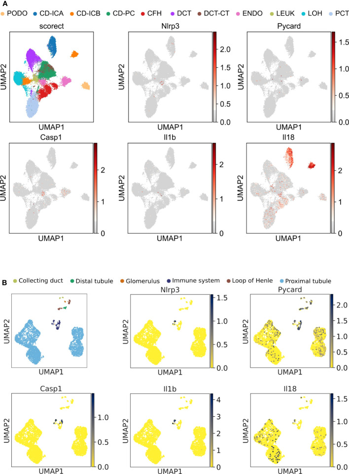
Background: The NLRP3 inflammasome integrates several danger signals into the activation of innate immunity and inflammation by secreting IL-1β and IL-18. Most published data relate to the NLRP3 inflammasome in immune cells, but some reports claim similar roles in parenchymal, namely epithelial, cells. For example, podocytes, epithelial cells critical for the maintenance of kidney filtration, have been reported to express NLRP3 and to release IL-β in diabetic kidney disease, contributing to filtration barrier dysfunction and kidney injury. We questioned this and hence performed independent verification experiments.

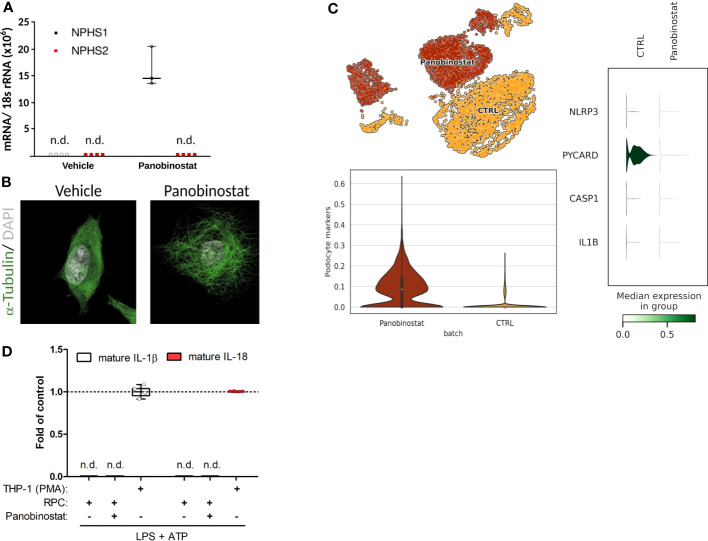
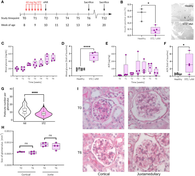
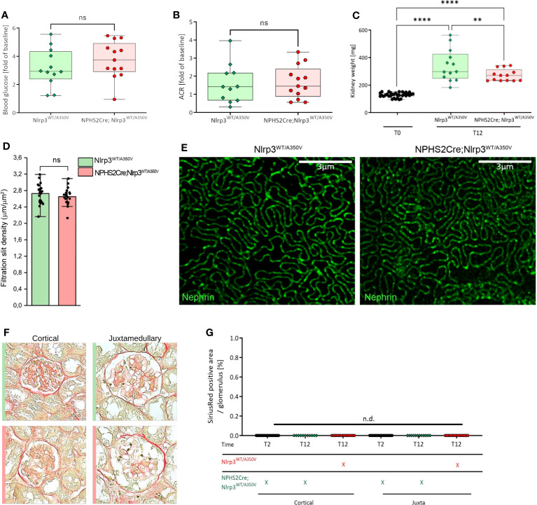
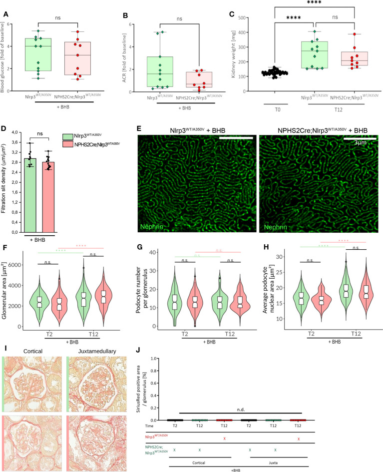
Methods: We studied the expression of inflammasome components in human and mouse kidneys and human podocytes using single-cell transcriptome analysis. Human podocytes were exposed to NLRP3 inflammasome agonists in vitro and we induced diabetes in mice with a podocyte-specific expression of the Muckle-Wells variant of NLRP3, leading to overactivation of the Nlrp3 inflammasome (Nphs2Cre;Nlrp3A350V) versus wildtype controls. Phenotype analysis included deep learning-based glomerular and podocyte morphometry, tissue clearing, and STED microscopy of the glomerular filtration barrier. The Nlrp3 inflammasome was blocked by feeding ß-hydroxy-butyrate.

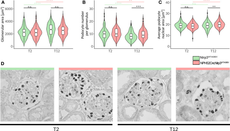
Results: Single-cell transcriptome analysis did not support relevant NLRP3 expression in parenchymal cells of the kidney. The same applied to primary human podocytes in which NLRP3 agonists did not induce IL-1β or IL-18 secretion. Diabetes induced identical glomerulomegaly in wildtype and Nphs2Cre;Nlrp3A350V mice but hyperfiltration-induced podocyte loss was attenuated and podocytes were larger in Nphs2Cre;Nlrp3A350V mice, an effect reversible with feeding the NLRP3 inflammasome antagonist ß-hydroxy-butyrate. Ultrastructural analysis of the slit diaphragm was genotype-independent hence albuminuria was identical.

Conclusion: Podocytes express low amounts of the NLRP3 inflammasome, if at all, and do not produce IL-1β and IL-18, not even upon introduction of the A350V Muckle-Wells NLRP3 variant and upon induction of podocyte stress. NLRP3-mediated glomerular inflammation is limited to immune cells.

Keywords: IL-1; chronic kidney disease; diabetes; inflammasome; inflammation; proteinuria.


Conflict of interest statement

The authors declare that the research was conducted in the absence of any commercial or financial relationships that could be construed as a potential conflict of interest.

Figures

Figure 1
Figure 1
Unbiased single-cell RNA sequencing of diabetic human and healthy murine kidneys indicates the absence of canonical NLRP3 inflammasome. (A) UMAP plot (upper left panel) and superimposed expression pattern of Nlrp3, Pycard, Casp1, IL1b, and IL18 of single nucleus RNA sequencing data from diabetic human kidneys (GSE131882). (B) Same analytic procedure as in (A), but for healthy C57BL/6N mice (GSE212273). PCT, proximal convoluted tubule; CFH, complement factor H; LOH, loop of Henle; DCT, distal convoluted tubule; CT, connecting tubule; CD, collecting duct; PC, principal cell; IC, intercalated cell; PODO, podocyte; ENDO, endothelium; MES, mesangial cell; and LEUK, leukocyte.
Figure 2
Figure 2
Primary human podocyte-like cells do not indicate complete functionality of canonical NLRP3 inflammasome. (A) Normalized NPHS1 and NPHS2 mRNA expression in primary human renal progenitor cells (hRPCs) after 48 h of vehicle or panobinostat treatment. (B) Confocal imaging of α-tubulin (green) and DAPI (white) stained vehicle and panobinostat treated hRPCs. (C) UMAP clustering for unbiased single-cell RNA sequencing data from vehicle (CTRL) or panobinostat-treated hRPCs (upper left panel, GSE195797). NLRP3, PYCARD, CASP1, and IL1B expression per UMAP cluster. (D) ELISAs of supernatants from panobinostat-treated hRPCs do not detect cleaved IL-1β and IL-18, respectively, after stimulation in contrast to phorbol myristate acetate treated THP-1 cells; n = 3, n.d., not detectable.
Figure 3
Figure 3
Female C57BL/6J mice respond with loss of β-cells, hyperglycemia, and albuminuria to streptozotocin (STZ) treatment and uninephrectomy (uNX). (A) Female C57BL6/J mice were injected with STZ (six intraperitoneal injections of 60 mg/kg BW every other day prior to uNX and followed for 4 and 10 weeks). (B) Immunohistochemistry for insulin in pancreatic tissue of control and STZ/uNX treated mice at T6. Time course (C, E) and T6 (D, F) of blood glucose (C, D) and albumin creatinine ratio (ACR; E, F) of STZ/uNX treated mice or control and STZ/uNX treated mice, respectively. (G) Podocyte number per glomerulus at T6. (H, I) Morphometric determination of the glomerular size of STZ/uNX treated mice from Periodic acid-Schiff (PAS) stained paraffin sections at indicated time points. n = 5, *p<0.05, ****p < 0.0001. ns, not significant.
Figure 4
Figure 4
Introduction of a podocyte-specific Nlrp3WT/A350V mutation does not aggravate the phenotype after STZ/uNX treatment. Female Nlrp3WT/A350V and Podo-Cre; Nlrp3WT/A350V mice underwent treatment indicated in Figure 4A . Blood glucose (A), ACR (B), and kidney weight (C) were determined at T12 (n ≥ 11). (D, E) Density of the glomerular slit diaphragm was investigated using stimulated emission depletion microscopy (STED) for nephrin-stained kidney sections of either genotype at T12 (n = 3). (F, G) Representative sirius red staining pictures quantified in (F) for signs of glomerulosclerosis.**p<0.01, ****p < 0.0001, n.d., not detectable. ns, not significant.
Figure 5
Figure 5
Deep learning-assisted morphometrics reveal podocyte hypertrophy in Podo-Cre; Nlrp3WT/A350V mice. A U-Net image segmentation algorithm was used to segment glomeruli and podocyte nuclei in WT-1-stained images (D) and calculate the glomerular area (A), average podocyte number per glomerulus (B), and average podocyte nuclear area (C). (D) Representative images of glomeruli from WT-1-stained kidney sections of female Nlrp3WT/A350V and Podo-Cre; Nlrp3WT/A350V mice after STZ and uNX at different time points. n ≥ 11, **p<0.01, ***p<0.001, ****p < 0.0001. n.s., not significant.
Figure 6
Figure 6
Treatment of Podo-Cre; Nlrp3WT/A350V mice with β-hydroxybutyrate (BHB) does rescue the Nlrp3WT/A350V induced phenotype. Female Nlrp3WT/A350V and Podo-Cre; Nlrp3WT/A350V mice underwent treatment indicated in Figure 3A and received BHB-enriched food ad libitum between T3 and T12. Blood glucose (A), ACR (B), and kidney weight (C) were determined at T12 (n ≥ 11). (D, E) Density of the glomerular slit diaphragm was investigated using stimulated emission depletion microscopy (STED) for nephrin-stained kidney sections of either genotype at T12 (n = 3). Deep learning assisted morphometrics to calculate the glomerular area (F), average podocyte number per glomerulus (G), and average podocyte nuclear area (H) from WT-1-stained kidney sections as shown in Figure 5 . Representative images of sirius red stained sections (I) quantified in (J) for signs of glomerulosclerosis. (n ≥ 11), ****p < 0.0001. ns, not significant.

Similar articles

Cited by

References

    1. Sharma BR, Kanneganti TD. NLRP3 inflammasome in cancer and metabolic diseases. Nat Immunol (2021) 22(5):550–9. doi: 10.1038/s41590-021-00886-5 - DOI - PMC - PubMed
    1. Chae JJ, Komarow HD, Cheng J, Wood G, Raben N, Liu PP, et al. . Targeted disruption of pyrin, the FMF protein, causes heightened sensitivity to endotoxin and a defect in macrophage apoptosis. Mol Cell (2003) 11(3):591–604. doi: 10.1016/s1097-2765(03)00056-x - DOI - PubMed
    1. Martinon F, Burns K, Tschopp J. The inflammasome: a molecular platform triggering activation of inflammatory caspases and processing of proIL-beta. Mol Cell (2002) 10(2):417–26. doi: 10.1016/s1097-2765(02)00599-3 - DOI - PubMed
    1. Zhao C, Zhao W. NLRP3 inflammasome-a key player in antiviral responses. Front Immunol (2020) 11:211. doi: 10.3389/fimmu.2020.00211 - DOI - PMC - PubMed
    1. Desai J, Steiger S, Anders HJ. Molecular pathophysiology of gout. Trends Mol Med (2017) 23(8):756–68. doi: 10.1016/j.molmed.2017.06.005 - DOI - PubMed

Publication types

LinkOut - more resources