

This is a preprint.
A Large Open Access Dataset of Brain Metastasis 3D Segmentations with Clinical and Imaging Feature Information
- PMID: 37744461
- PMCID: PMC10516117
A Large Open Access Dataset of Brain Metastasis 3D Segmentations with Clinical and Imaging Feature Information
Update in
-
A large open access dataset of brain metastasis 3D segmentations on MRI with clinical and imaging information.Sci Data. 2024 Feb 29;11(1):254. doi: 10.1038/s41597-024-03021-9. Sci Data. 2024. PMID: 38424079 Free PMC article.
Abstract
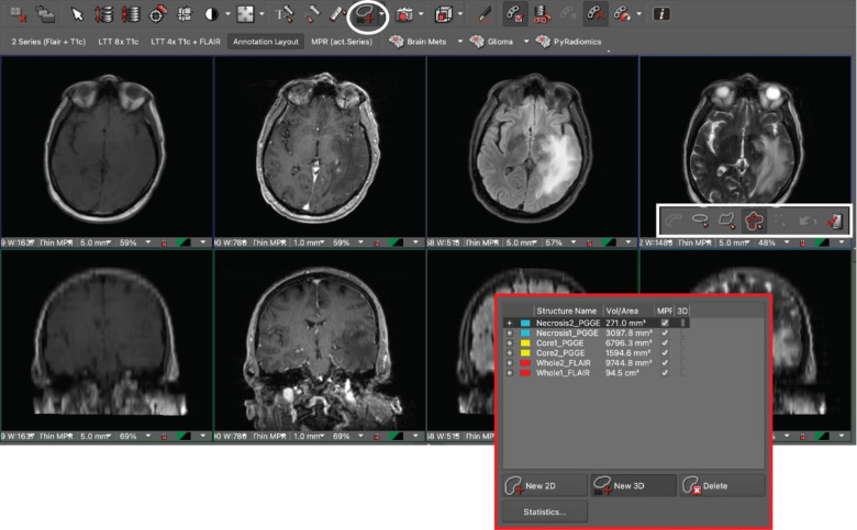
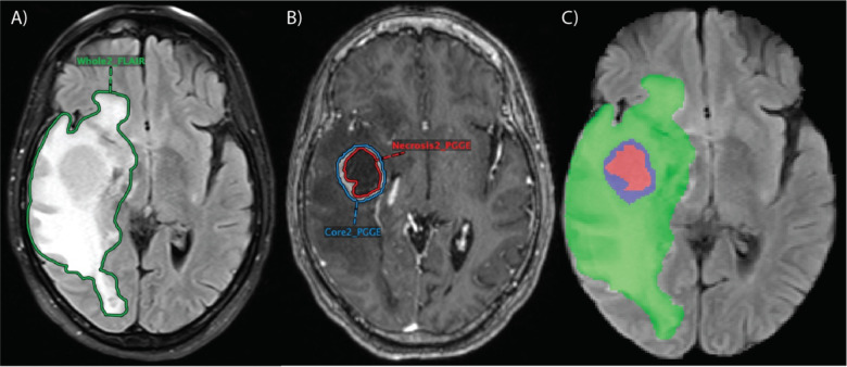
Resection and whole brain radiotherapy (WBRT) are the standards of care for the treatment of patients with brain metastases (BM) but are often associated with cognitive side effects. Stereotactic radiosurgery (SRS) involves a more targeted treatment approach and has been shown to avoid the side effects associated with WBRT. However, SRS requires precise identification and delineation of BM. While many AI algorithms have been developed for this purpose, their clinical adoption has been limited due to poor model performance in the clinical setting. Major reasons for non-generalizable algorithms are the limitations in the datasets used for training the AI network. The purpose of this study was to create a large, heterogenous, annotated BM dataset for training and validation of AI models to improve generalizability. We present a BM dataset of 200 patients with pretreatment T1, T1 post-contrast, T2, and FLAIR MR images. The dataset includes contrast-enhancing and necrotic 3D segmentations on T1 post-contrast and whole tumor (including peritumoral edema) 3D segmentations on FLAIR. Our dataset contains 975 contrast-enhancing lesions, many of which are sub centimeter, along with clinical and imaging feature information. We used a streamlined approach to database-building leveraging a PACS-integrated segmentation workflow.
Conflict of interest statement
M.S.A. has collaborations with Visage Imaging, Inc., Blue Earth Diagnostics, Telix, and AAA. She also has a KL2 TR00186 grant from the NCATS foundation. M.L. is an employee and stockholder of Visage Imaging, Inc., and unrelated to this work, receives funding from NIH/NCI R01 CA206180 and NIH/NCI R01 CA275188. W.H. and M.W. are employees and stockholders of Visage Imaging GmbH. K.B. is an employee of Visage Imaging GmbH. C.K. receives royalties from Primal Pictures 3D Informa, has grant funding from the NIH, and has received the Core Curriculum grant from the American Society of Head and Neck Radiology, all unrelated to this work. The remaining co-authors do not have any competing interests.
Figures
References
Publication types
Grants and funding
LinkOut - more resources
Full Text Sources