

Comparative study in vivo of the osseointegration of 3D-printed and plasma-coated titanium implants
- PMID: 37744721
- PMCID: PMC10514715
- DOI: 10.5312/wjo.v14.i9.682
Comparative study in vivo of the osseointegration of 3D-printed and plasma-coated titanium implants
Abstract
Background: Total hip arthroplasty is a common surgical treatment for elderly patients with osteoporosis, particularly in postmenopausal women. In such cases, highly porous acetabular components are a favorable option in achieving osseointegration. However, further discussion is needed if use of such acetabular components is justified under the condition of normal bone mass.
Aim: To determine the features of osseointegration of two different types of titanium implants [3-dimensional (3D)-printed and plasma-coated titanium implants] in bone tissue of a distal metaphysis in a rat femur model.
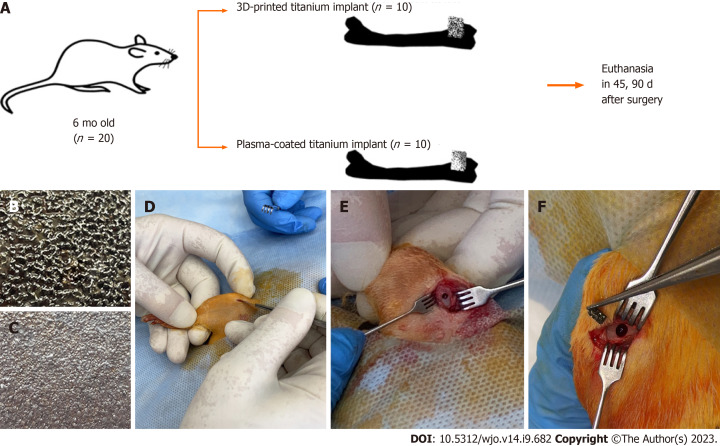
Methods: This study was performed on 20 white male laboratory rats weighing 300-350 g aged 6 mo. Rats were divided into two groups of 10 animals, which had two different types of implants were inserted into a hole defect (2 × 3 mm) in the distal metaphysis of the femur: Group I: 3D-printed titanium implant (highly porous); Group II: Plasma-coated titanium implant. After 45 and 90 d following surgery, the rats were sacrificed, and their implanted femurs were extracted for histological examination. The relative perimeter (%) of bone trabeculae [bone-implant contact (BIC%)] and bone marrow surrounding the titanium implants was measured.
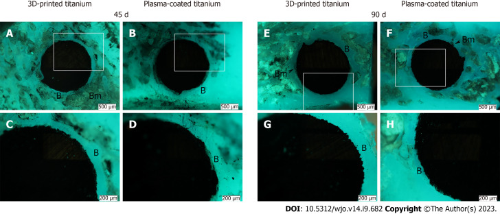
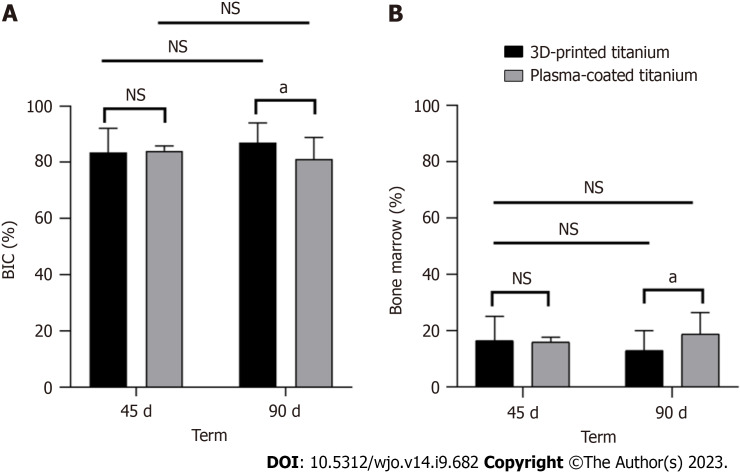
Results: Trabecular bone tissue was formed on the 45th day after implantation around the implants regardless of their type. 45 d after surgery, group I (3D-printed titanium implant) and group II (plasma-coated titanium implant) did not differ in BIC% (83.51 ± 8.5 vs 84.12 ± 1 .73; P = 0.838). After 90 d, the BIC% was higher in group I (87.04 ± 6.99 vs 81.24 ± 7.62; P = 0.049), compared to group II. The relative perimeter of the bone marrow after 45 d did not differ between groups and was 16.49% ± 8.58% for group I, and 15.88% ± 1.73% for group II. Futhermore, after 90 d, in group I the relative perimeter of bone marrow was 1.4 times smaller (12.96 ± 6.99 vs 18.76 ± 7.62; P = 0.049) compared to the relative perimeter of bone marrow in group II.
Conclusion: The use of a highly porous titanium implant, manufactured with 3D printing, for acetabular components provides increased osseointegration compared to a plasma-coated titanium implant.
Keywords: 3-dimensional printing; Femur; Hip arthroplasty; Microscopy; Porosity; Rats.
©The Author(s) 2023. Published by Baishideng Publishing Group Inc. All rights reserved.
Conflict of interest statement
Conflict-of-interest statement: The authors declare that they have no conflict of interest.
Figures
Similar articles
-
Comparative analysis of osseointegration in various types of acetabular implant materials.Hip Int. 2018 Nov;28(6):622-628. doi: 10.1177/1120700018759314. Epub 2018 May 9. Hip Int. 2018. PMID: 29742946
-
Implant-delivered Alendronate Causes a Dose-dependent Response on Net Bone Formation Around Porous Titanium Implants in Canines.Clin Orthop Relat Res. 2016 May;474(5):1224-33. doi: 10.1007/s11999-016-4714-6. Epub 2016 Feb 1. Clin Orthop Relat Res. 2016. PMID: 26831478 Free PMC article.
-
3D laser-printed porous Ti6Al4V dental implants for compromised bone support.J Formos Med Assoc. 2020 Jan;119(1 Pt 3):420-429. doi: 10.1016/j.jfma.2019.07.023. Epub 2019 Aug 3. J Formos Med Assoc. 2020. PMID: 31387841
-
Advanced Surface Modification for 3D-Printed Titanium Alloy Implant Interface Functionalization.Front Bioeng Biotechnol. 2022 Mar 1;10:850110. doi: 10.3389/fbioe.2022.850110. eCollection 2022. Front Bioeng Biotechnol. 2022. PMID: 35299643 Free PMC article. Review.
-
Characterisation of 3D-printed acetabular hip implants.EFORT Open Rev. 2024 Sep 2;9(9):862-872. doi: 10.1530/EOR-23-0182. EFORT Open Rev. 2024. PMID: 39222334 Free PMC article. Review.
Cited by
-
Applying 3D-printed prostheses to reconstruct critical-sized bone defects of tibial diaphysis (> 10 cm) caused by osteomyelitis and aseptic non-union.J Orthop Surg Res. 2024 Jul 20;19(1):418. doi: 10.1186/s13018-024-04926-2. J Orthop Surg Res. 2024. PMID: 39033286 Free PMC article.
-
Synthetic Nanopillars for Stimulating Osteoblast Activity and Osteointegration in Bone-Related Disorders.Int J Nanomedicine. 2025 Feb 19;20:2205-2223. doi: 10.2147/IJN.S501963. eCollection 2025. Int J Nanomedicine. 2025. PMID: 39990287 Free PMC article. Review.
References
-
- Dall’Ava L, Hothi H, Di Laura A, Henckel J, Hart A. 3D printed acetabular cups for total hip arthroplasty: A review article. Metals (Basel) 2019;9:729.
-
- Malahias MA, Kostretzis L, Greenberg A, Nikolaou VS, Atrey A, Sculco PK. Highly Porous Titanium Acetabular Components in Primary and Revision Total Hip Arthroplasty: A Systematic Review. J Arthroplasty. 2020;35:1737–1749. - PubMed
-
- Small SR, Berend ME, Howard LA, Rogge RD, Buckley CA, Ritter MA. High initial stability in porous titanium acetabular cups: a biomechanical study. J Arthroplasty. 2013;28:510–516. - PubMed
LinkOut - more resources
Full Text Sources
