

Isolated Flat Epithelial Atypia: Upgrade Outcomes After Multidisciplinary Review-Based Management Using Excision or Imaging Surveillance
- PMID: 37744722
- PMCID: PMC10516722
- DOI: 10.1093/jbi/wbad049
Isolated Flat Epithelial Atypia: Upgrade Outcomes After Multidisciplinary Review-Based Management Using Excision or Imaging Surveillance
Abstract
Objective: To compare flat epithelial atypia (FEA) upgrade rates after excision versus surveillance and to identify variables associated with upgrade.
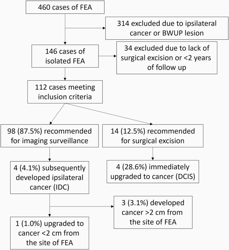
Methods: This single-institution retrospective study identified isolated FEA cases determined by percutaneous biopsy from April 2005 through July 2022 with excision or ≥2 years surveillance. All cases were recommended for excision or surveillance based on multidisciplinary discussion of clinical, imaging, and pathologic variables with emphasis on sampling adequacy and significant atypia. Truth was determined by pathology at excision or the absence of cancer on surveillance. Upgrade was defined as cancer occurring ≤2 cm from the biopsy site. Demographic, imaging, and biopsy variables were compared between those that did and did not upgrade.
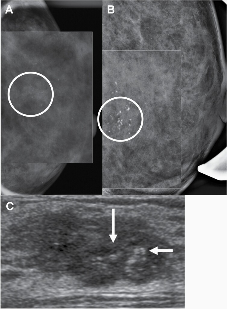
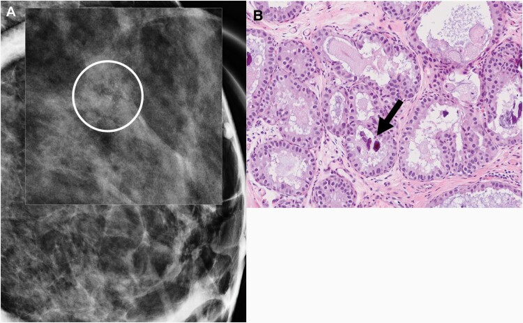
Results: Among 112 cases of isolated FEA, imaging findings included calcifications in 81.3% (91/112), MRI lesions in 11.6% (13/112), and distortions or masses in 7.1% (8/112). Excision was recommended in 12.5% (14/112) and surveillance in 87.5% (98/112) of cases. Among those recommended for excision, 28.6% (4/14) of cases were upgraded, all to ductal carcinoma in situ. In those recommended for surveillance, 1.0% (1/98) were upgraded to invasive cancer. Overall, FEA had a 4.5% (5/112) upgrade rate, and 2.7% (3/112) also developed cancer >2 cm from the FEA. There were no significant differences in demographic, imaging, and biopsy variables between those that did and did not upgrade to cancer.
Conclusion: Multidisciplinary management of isolated FEA distinguishes those at higher risk of upgrade to cancer (28.6%) in whom surgery is warranted from those at low risk of upgrade (1.0%) who can be managed non-operatively.
Keywords: benign with upgrade potential; flat epithelial atypia; multidisciplinary review.
© Society of Breast Imaging 2023. All rights reserved. For permissions, please e-mail: journals.permissions@oup.com.
Conflict of interest statement
G.J.W. is a consultant for Siemens and an editor for UpToDate. The remaining authors have no conflicts of interest declared.
Figures


References
-
- Tan PH, Ellis I, Allison K, et al. ; WHO Classification of Tumours Editorial Board. The 2019 World Health Organization classification of tumours of the breast. Histopathology 2020;77(2):181–185. - PubMed
-
- Batohi B, Fang C, Michell MJ, et al. An audit of mammographic screen detected lesions of uncertain malignant potential (B3) diagnosed on initial image guided needle biopsy: how has our practice changed over 10 years? Clin Radiol 2019;74(8):653.e19–653.e25. - PubMed
-
- D’Orsi CJ, Sickles EA, Mendelson EB, Morris EA, et al. ACR BI-RADS ® Atlas, Breast Imaging Reporting and Data System . Reston, VA: American College of Radiology; 2013.
-
- Aulmann S, Braun L, Mietzsch F, et al. Transitions between flat epithelial atypia and low-grade ductal carcinoma in situ of the breast. Am J Surg Pathol 2012;36(8):1247–1252. - PubMed
Grants and funding
LinkOut - more resources
Full Text Sources
Miscellaneous