Review
. 2023 Sep 8;8(37):33202-33228.
doi: 10.1021/acsomega.3c03746. eCollection 2023 Sep 19.

Cyclodextrin Host-Guest Recognition in Glucose-Monitoring Sensors

Affiliations
Review

Cyclodextrin Host-Guest Recognition in Glucose-Monitoring Sensors

Siamak Javanbakht et al. ACS Omega. .

Abstract

Diabetes mellitus is a prevalent chronic health condition that has caused millions of deaths worldwide. Monitoring blood glucose levels is crucial in diabetes management, aiding in clinical decision making and reducing the incidence of hypoglycemic episodes, thereby decreasing morbidity and mortality rates. Despite advancements in glucose monitoring (GM), the development of noninvasive, rapid, accurate, sensitive, selective, and stable systems for continuous monitoring remains a challenge. Addressing these challenges is critical to improving the clinical utility of GM technologies in diabetes management. In this concept, cyclodextrins (CDs) can be instrumental in the development of GM systems due to their high supramolecular recognition capabilities based on the host-guest interaction. The introduction of CDs into GM systems not only impacts the sensitivity, selectivity, and detection limit of the monitoring process but also improves biocompatibility and stability. These findings motivated the current review to provide a comprehensive summary of CD-based blood glucose sensors and their chemistry of glucose detection, efficiency, and accuracy. We categorize CD-based sensors into four groups based on their modification strategies, including CD-modified boronic acid, CD-modified mediators, CD-modified nanoparticles, and CD-modified functionalized polymers. These findings shed light on the potential of CD-based sensors as a promising tool for continuous GM in diabetes mellitus management.


Conflict of interest statement

The authors declare no competing financial interest.

Figures

Figure 1
Figure 1
Chemical structures and cartoon representations of α-, β-, and γ-cyclodextrin.
Figure 2
Figure 2
Various glucose-monitoring systems based on cyclodextrins.
Figure 3
Figure 3
Schematic illustration of the boronic acid fluorophore/β-CD complex response mechanism for sugar binding.
Figure 4
Figure 4
Proposed schematic illustration of (A) the structures of inclusion complexes of pyrene-based fluorescent probe/γ-CD with (a) fructose and (b) glucose and (B) structures of the inclusion complex of anthracene-based fluorescent probes (a) with/without sugar, (b) with fructose/mannose, and (c) with glucose/galactose.
Figure 5
Figure 5
Proposed schematic illustration of (A) the formed 2:1 inclusion complex of BA-Azo/γ-CD in the presence of d-glucose, (B) the formed inclusion complexes of the 3-fluorophenylboronic-acid-based anthracene-type probe /γ-CD, and (C) the sugar recognition with the C1-APB/3-PB-γ-CD, (a) fructose, and (b) glucose.
Figure 6
Figure 6
Proposed schematic illustration of the interaction of a 2:2 STDBA−γ-CD inclusion complex with fructose and glucose.
Figure 7
Figure 7
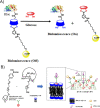
(A) Schematic representation of the glucose sensor based on bioluminescence quenching. (B) Schematic representation of the PBA-AD that is attached to the β-CD amphiphile (β-CDA) bilayer construction in aqueous solution via host–guest interactions and the competitive binding with ARS.
Figure 8
Figure 8
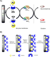
(A) Schematic representation of the glucose sensing mechanism for the modified platinum electrode surface by GNPs/CD-Fc/GOx. (B) The preparation of the Au-CMCD and Au-CMCD/PCMCD electrodes and current changes of the electrodes to (1) 0.1 mM glucose and 0.1 mM glucose in the presence of 0.1 mM (2) dopamine, (3) tyrosine, (4) guanine, (5) thymine, (6) folic acid, (7) glycerol, (8) ascorbic acid, (9) uric acid, (10) chloramphenicol, (11) fructose, (12) galactose, and (13) mannose, respectively.
Figure 9
Figure 9
(A) Schematic illustration of the UC-FRET glucose biosensor mechanism. Reprinted with permission from ref (95). Copyright 2011 Elsevier. (B) Schematic illustration of the GOx-ADA/CD-PAMAM/PtNP/Au-based glucose biosensor electrode fabrication.
Figure 10
Figure 10
(A) (a) Schematic representation of the β-CD/CuNP synthesis and (b) colorimetric detection schematic for H2O2 and glucose. (B) Schematic representation of the GM mechanism by a fluorescent FRET system between 3-hydroxyflavone and β-CD/ZnS-QDs.
Figure 11
Figure 11
(A) Selectivity of N- and Fe-CQDs in the presence of different compounds and (B) the comparison of their absorbance intensity. Reprinted with permission from ref (103). Copyright © 2020. Yan Li et al. Journal of Nanomaterials.
Figure 12
Figure 12
Illustration of a β-CD-based FRET glucose-monitoring system.
Figure 13
Figure 13
(A) Probable mechanism for chemiluminescence (CL) enhancement of the luminol–H2O2 system by MOF-235/β-CD hybrids. (B) The linear relationship between the concentration of glucose and net CL intensity under the optimized conditions. (C) Selectivity test for glucose detection by considering the relative CL intensity. Reprinted with permission from ref (105). Copyright 2018 Elsevier.
Figure 14
Figure 14
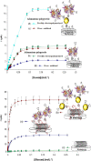
Structure of the prepared SWCNT electrodes and related standard calibration curves for glucose (applied potential of 0.7 V vs SCE, 0.1 M phosphate buffer, pH 7.0). Reprinted with permission from ref (106). Copyright 2009 Elsevier.
Figure 15
Figure 15
(A) Schematic illustration of the GOx/β-CD/MWCNTs/GCE fabrication. Reprinted with permission from ref (150). Copyright 2022 Elsevier. (B) Preparation of the rGO/CD composite as a glucose biosensor (a). The CV response of modified electrodes with blank CD (b-a), CD/GOx (b-b), rGO/CD (b-c), and rGO/CD/GOx (b-d) in the scanning range from −0.7 to 0 V. Reprinted with permission from ref. Copyright 2015 Wiley-VCH.
Figure 16
Figure 16
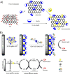
(A) Schematic represents the synthetic strategies of the graphene-based metal nanocomposite via in situ growth and post immobilization. (B) Schematic representation of the preparation procedure of β-CD-AgNPs-rGO, FcCA-CM-β-CD, GOx-FcCA-CM-β-CD, and GOxFcCD/AgNPs@rGO/GCE and a dual-path electron transfer mechanism for the modified electrochemical glucose biosensor.
Figure 17
Figure 17
Schematic illustration of the preparation of the PVA/β-CD/CA/GOx hydrogel and its application on an epidermal patch with an iontophoresis mechanism.
Figure 18
Figure 18
Schematic illustration of (A) the iontophoresis printable cellulose/β-CD/GOx NF electrode and (B) its iontophoresis mechanism on the epidermal glucose detection. Reprinted with permission from ref (120). Copyright 2019. Kyu Oh Kim et al. Royal Society of Chemistry.
Figure 19
Figure 19
(A) Schematic of the patch-type glucose sensor employing PVA/BTCA/β-CD/GOx/AuNP NF hydrogels on electrodes and the mechanism of glucose sensing for the noninvasive real-time monitoring of glucose in sweat. (B) Schematic of the preparation of the PVA/BTCA/β-CD/GOx/AuNP complex dope solution for electrospinning. (C) Transparent PVA/BTCA/β-CD/GOx/AuNP nanofibrous hydrogel process (containing optical and SEM images). Reprinted with permission from ref (121). Copyright 2020. Gun Jin Kim et al., Pub Med.

References

    1. Guo X.; Li H.; Xu H.; Woo S.; Dong H.; Lu F.; Lange A. J.; Wu C. Glycolysis in the control of blood glucose homeostasis. Acta Pharm. Sin B 2012, 2 (4), 358–367. 10.1016/j.apsb.2012.06.002. - DOI
    1. Panlasigui L. N.; Thompson L. U. Blood glucose lowering effects of brown rice in normal and diabetic subjects. Int. J. Food Sci. Nutr 2006, 57 (3–4), 151–158. 10.1080/09637480500410879. - DOI - PubMed
    1. Saberzadeh-Ardestani B.; Karamzadeh R.; Basiri M.; Hajizadeh-Saffar E.; Farhadi A.; Shapiro A. M. J.; Tahamtani Y.; Baharvand H. Type 1 Diabetes Mellitus: Cellular and Molecular Pathophysiology at A Glance. Cell J. 2018, 20 (3), 294–301. 10.22074/cellj.2018.5513. - DOI - PMC - PubMed
    1. Ozougwu J.; Obimba K.; Belonwu C.; Unakalamba C. The pathogenesis and pathophysiology of type 1 and type 2 diabetes mellitus. J. Physiol Pathophysiol 2013, 4 (4), 46–57. 10.5897/JPAP2013.0001. - DOI
    1. Galicia-Garcia U.; Benito-Vicente A.; Jebari S.; Larrea-Sebal A.; Siddiqi H.; Uribe K. B.; Ostolaza H.; Martin C. Pathophysiology of Type 2 Diabetes Mellitus. Int. J. Mol. Sci. 2020, 21 (17), 6275.10.3390/ijms21176275. - DOI - PMC - PubMed