. 2023 Sep 5:14:1190530.
doi: 10.3389/fmicb.2023.1190530. eCollection 2023.

Phenotypic assay for cytotoxicity assessment of Balamuthia mandrillaris against human neurospheroids

Affiliations

Phenotypic assay for cytotoxicity assessment of Balamuthia mandrillaris against human neurospheroids

Narisara Whangviboonkij et al. Front Microbiol. .

Abstract

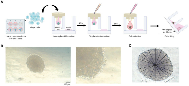
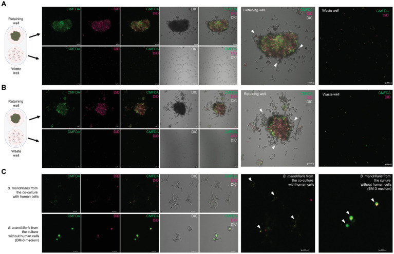
Introduction: The phenotypic screening of drugs against Balamuthia mandrillaris, a neuropathogenic amoeba, involves two simultaneous phases: an initial step to test amoebicidal activity followed by an assay for cytotoxicity to host cells. The emergence of three-dimensional (3D) cell cultures has provided a more physiologically relevant model than traditional 2D cell culture for studying the pathogenicity of B. mandrillaris. However, the measurement of ATP, a critical indicator of cell viability, is complicated by the overgrowth of B. mandrillaris in coculture with host cells during drug screening, making it challenging to differentiate between amoebicidal activity and drug toxicity to human cells.

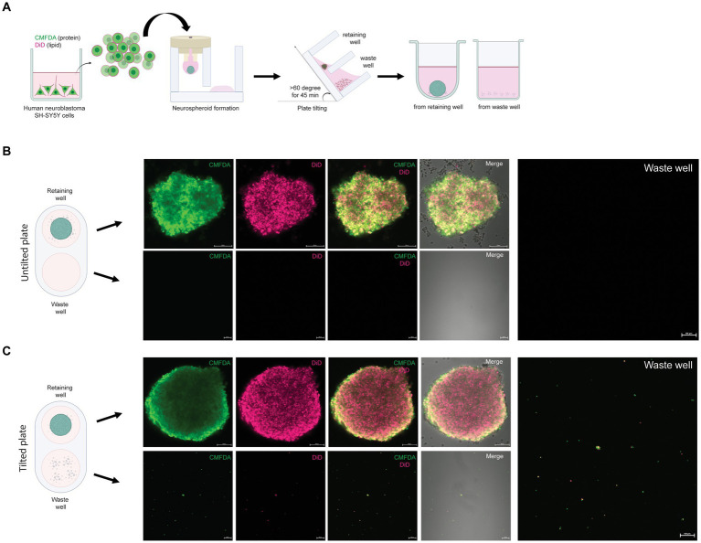
Methods: To address this limitation, we introduce a novel assay that utilizes three-dimensional hanging spheroid plates (3DHSPs) to evaluate both activities simultaneously on a single platform.

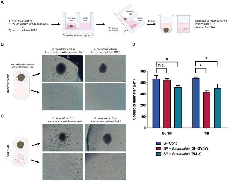
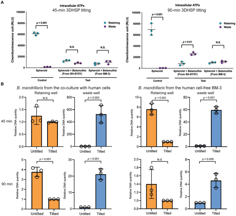
Results and discussion: Our study showed that the incubation of neurospheroids with clinically isolated B. mandrillaris trophozoites resulted in a loss of neurospheroid integrity, while the ATP levels in the neurospheroids decreased over time, indicating decreased host cell viability. Conversely, ATP levels in isolated trophozoites increased, indicating active parasite metabolism. Our findings suggest that the 3DHSP-based assay can serve as an endpoint for the phenotypic screening of drugs against B. mandrillaris, providing a more efficient and accurate approach for evaluating both parasite cytotoxicity and viability.

Keywords: Balamuthia mandrillaris; cytotoxicity; drug discovery; granulomatous amoebic encephalitis; neglected disease; neurospheroid; tropical disease.


Conflict of interest statement

The authors declare that the research was conducted in the absence of any commercial or financial relationships that could be construed as a potential conflict of interest.

Figures

Figure 1
Figure 1
Neurospheroid formation in a three-dimensional hanging spheroid plate (3DHSP). (A) Schematic diagram of neurospheroid formation in the 3DHSP. Single cells were hung in a drop in the retaining well, while the waste well was filled with the culture medium. After a 48-h incubation, B. mandrillaris trophozoites were incubated with neurospheroids. After 24 h of trophozoite incubation, cells in the hanging drop were collected by adding culture medium. The 3DHSP was tilted at an angle of 60°C for 45 min to separate the trophozoites from the neurospheroids. (B) Representative images of human neurospheroids in after 48 h of hanging drop culture. A higher magnification is shown on the right-hand side (scale bar = 100 μm). The dark hypoxic area is marked by the white line. (C) Measurement of spheroid diameter. ImageJ was used to measure the diameter. Due to incomplete spherical shape, ten lines are drawn across the neurospheroid, and the lengths of the drawn lines were averaged.
Figure 2
Figure 2
Removal of single SH-SY5Y cells from the neurospheroid. (A) Illustration of the method for forming fluorescent neurospheroids. Human neuroblastoma SH-SY5Y cells were incubated with protein-binding CMFDA and lipid-binding DiD in the 2D cell culture well. After cell labeling, single SH-SY5Y cells were subjected to neurospheroid formation in the 3DHSP. After plate tilting, the neurospheroids and the cells in the waste well were transferred to a round or flat bottom-containing plate, respectively. The fluorescence-labeled neurospheroids and cells were imaged under a confocal microscope. (B) Confocal microscopic images of SH-SY5Y neurospheroids in the retaining well (upper panel, scale bar = 100 μm) and waste well (lower panel, scale bar = 100 μm) of the untilted plate. Images of CMFDA-and DiD-labeled neurospheroid are merged with the differential interference contrast (Merge). A higher magnification of the CMFDA-and DiD-visualizing waste well is shown on the right-hand side (scale bar = 100 μm). (C) Confocal microscopic images of SH-SY5Y neurospheroids in the retaining well (upper panel, scale bar = 100 μm) and the waste well (lower panel, scale bar = 100 μm) after tilting for 90 min. Images of CMFDA-and DiD-labeled neurospheroid are merged with the differential interference contrast (Merge). A higher magnification of the CMFDA-and DiD-visualizing waste well is shown on the right-hand side (scale bar = 100 μm).
Figure 3
Figure 3
The effects of B. mandrillaris trophozoites on the integrity of neurospheroids. (A) Schematic diagram illustrating the assessment of the cytotoxicity of trophozoites against neurospheroids. The trophozoites were obtained from two sources: the coculture with human SH-SY5Y cells and the human cell-free BM-3 culture. Following 3DHSP tilting, the neurospheroids and cells in the waste well were transferred into a new round bottom and flat bottom plate for measuring the intracellular ATP and neurospheroid size. (B) Illustration of neurospheroids cultured with and without B. mandrillaris trophozoites in the hanging drops of the untilted plate. The spherical shape of the neurospheroid in the drop observed under a stereomicroscope. Scale bar, 100 μm. (C) Illustration of the neurospheroid and B. mandrillaris trophozoites in the retaining well and the waste well after plate tilting. (D) Sizes of the neurospheroids in the retaining wells were calculated from the untilted and tilted plates. Due to asymmetry, several lines of diameter were drawn and subjected to the calculation of spheroid diameter using ImageJ (left panel). The bar graph is the diameter of the neurospheroid (right panel). The B. mandrillaris trophozoites used in the coculture with neurospheroid (SP) were from the culture with human neuroblastoma cells (SH-SY5Y) or the feeder-free culture (BM-3).
Figure 4
Figure 4
The effects of B. mandrillaris trophozoites on the survival of neurospheroids. (A) The level of intracellular ATP produced by the neurospheroid in the retaining wells and the B. mandrillaris trophozoites in the waste wells. The control group consisted of human neurospheroids without trophozoites, while the test groups consisted of human neurospheroids cocultured with trophozoites. The 3DHSPs were tilted for 45 and 90 min (left and right panels, respectively). Each colored dot represents three biological replicates. (B) PCR data show the relative DNA quantity of B. mandrillaris DNA remaining in the retaining and waste wells before and after 3DHSP tilting for 45 and 90 min (upper and lower panel, respectively). Trophozoites were obtained from a culture with human neuroblastoma SH-SY5Y cells (left panel) and a feeder-free BM-3 medium (right panel). Circles represent triplicate wells of the quantitative PCR.
Figure 5
Figure 5
Cytophagy of the B. mandrillaris trophozoites in the 3DHSP. (A) Representative images of human neurospheroids cocultured with B. mandrillaris trophozoites obtained from culture with human neuroblastoma SH-SY5Y cells. Scale bars, 100 μm. (B) Representative images of human neurospheroids cocultured with B. mandrillaris trophozoites obtained from the feeder-free culture (BM-3 medium). Scale bars, 100 μm. (C) Zoomed-in images of B. mandrillaris trophozoites. The upper panels are representative images of the trophozoites obtained from (A), while the lower panels are images of the trophozoites obtained from (B). Microscopic images were captured in differential interference contrast (DIC) and fluorescence modes. The 3DHSP was tilted for 90 min. Scale bars, 20 μm. CMFDA, the protein-binding fluorophore, and DiD, the lipid-binding fluorophore.

Similar articles

Cited by

References

    1. Anada T., Fukuda J., Sai Y., Suzuki O. (2012). An oxygen-permeable spheroid culture system for the prevention of central hypoxia and necrosis of spheroids. Biomaterials 33, 8430–8441. doi: 10.1016/j.biomaterials.2012.08.040, PMID: - DOI - PubMed
    1. Bassler B. L., Losick R. (2006). Bacterially speaking. Cells 125, 237–246. doi: 10.1016/j.cell.2006.04.001, PMID: - DOI - PubMed
    1. Biedler J. L., Roffler-Tarlov S., Schachner M., Freedman L. S. (1978). Multiple neurotransmitter synthesis by human neuroblastoma cell lines and clones. Cancer Res. 38, 3751–3757. PMID: - PubMed
    1. Cangelosi G. A., Meschke J. S. (2014). Dead or alive: molecular assessment of microbial viability. Appl. Environ. Microbiol. 80, 5884–5891. doi: 10.1128/AEM.01763-14, PMID: - DOI - PMC - PubMed
    1. Chan G. K., Kleinheinz T. L., Peterson D., Moffat J. G. (2013). A simple high-content cell cycle assay reveals frequent discrepancies between cell number and ATP and MTS proliferation assays. PLoS One 8:e63583. doi: 10.1371/journal.pone.0063583, PMID: - DOI - PMC - PubMed

LinkOut - more resources