. 2023 Sep 8:10:1188105.
doi: 10.3389/fvets.2023.1188105. eCollection 2023.

Viral skin diseases in odontocete cetaceans: gross, histopathological, and molecular characterization of selected pathogens

Affiliations

Viral skin diseases in odontocete cetaceans: gross, histopathological, and molecular characterization of selected pathogens

Simone Segura-Göthlin et al. Front Vet Sci. .

Abstract

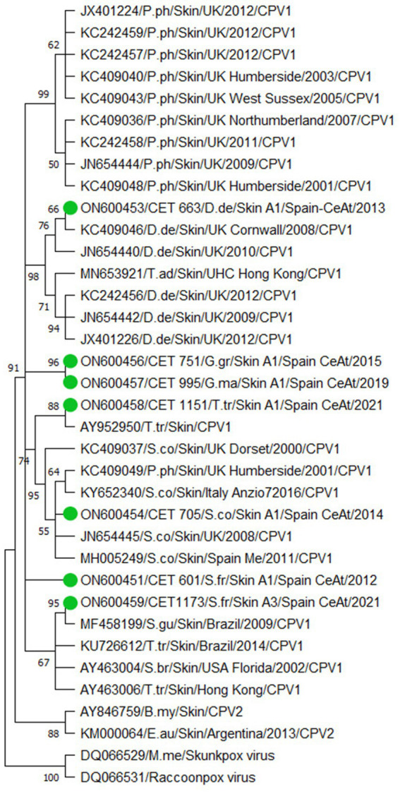
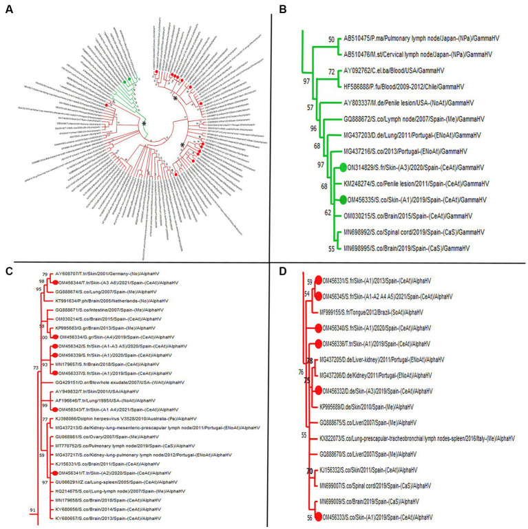
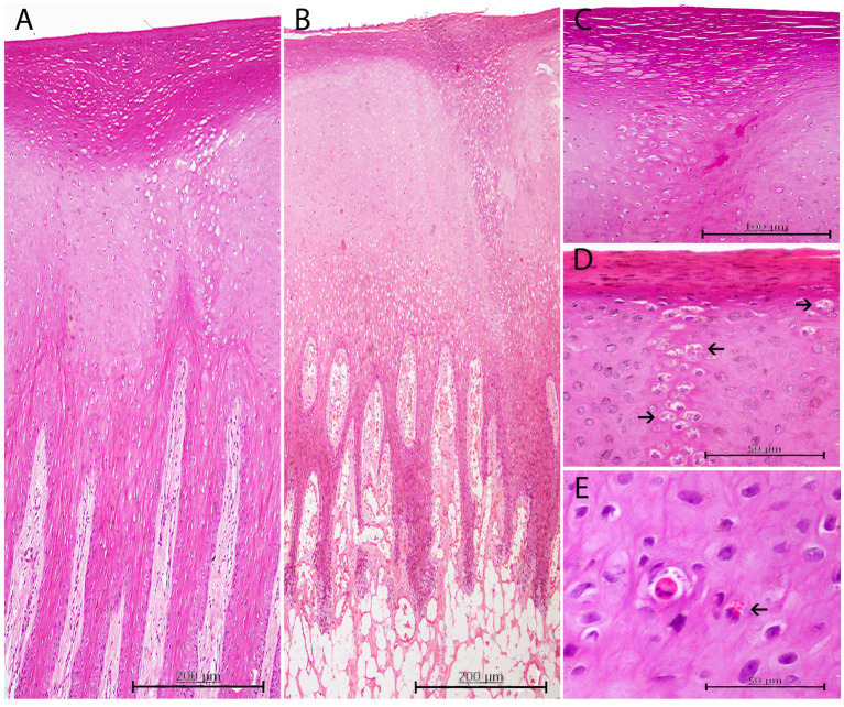
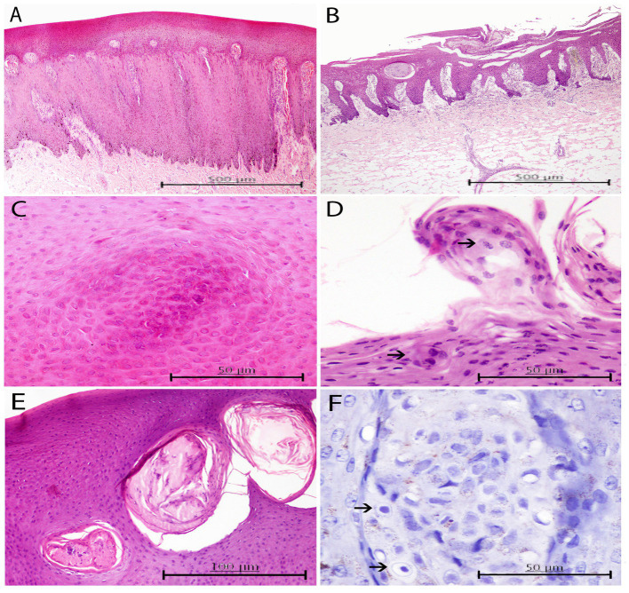
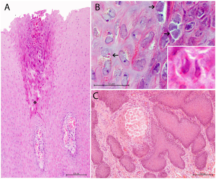
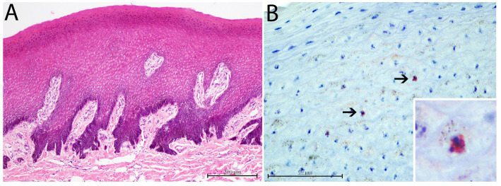
Fifty-five skin lesions from 31 stranded cetaceans along the Canary coasts (2011-2021) were submitted to macroscopic, histological, and molecular analyses to confirm infection by cetacean poxvirus, herpesvirus and cetacean morbillivirus. They were macroscopically categorized into eight categories with respective subcategories according to their color, shape, size, and consistency. Cetacean poxvirus was detected in 54.54% of the skin lesions through real-time and conventional PCRs based on the DNA polymerase gene. Additionally, herpesvirus and morbillivirus were currently detected from 43.63 and 1.82% of the cutaneous lesions, respectively. Coinfection of poxvirus and herpesvirus was detected in nine of them (16.36%), which makes the present study the first to report coinfection by both pathogens in skin lesions in cetaceans. A plausible approach to histopathological characterization of poxvirus-and herpesvirus-positive skin lesions was established. Hyperkeratosis, acanthosis, ballooning degeneration, and intracytoplasmic inclusion bodies in vacuolized keratinocytes through the stratum spinosum were common findings in poxvirus skin lesions. Alphaherpesvirus was associated with a prominent acanthotic epidermis, moderate necrosis, multifocal dyskeratosis, and irregular keratinocytes with both cellular and nuclei pleomorphism. The common histopathological findings of both pathogens were observed in coinfection lesions. However, those associated with herpesvirus were considerably more remarkable. Relationships between molecular and microscopic findings were observed for the lesions that showed tattoo-like and tortuous patterns. Further multidisciplinary diagnostic studies of infected skin lesions are needed to understand the epidemiology of these emerging infectious diseases.

Keywords: cetacean poxvirus; coinfection; herpesvirus; histopathology; molecular diagnosis; morbillivirus; skin lesions.


Conflict of interest statement

The authors declare that the research was conducted in the absence of any commercial or financial relationships that could be construed as a potential conflict of interest.

Figures

Figure 1
Figure 1
Phylogenetic analysis based on 29 nucleotide sequences from the polymerase gene of cetacean poxvirus. Seven sequences obtained from this study are denoted in colored green circles. The accession number, the identification number, the host, the geographic stranding, and the date of collection were used to identify the nucleotide sequences. B.my (Balaena mysticetus); D.de (Delphinus delphis); E.au (Eubalaena australis); G.gr (Grampus griseus); G.ma (Globicephala macrorhynchus); M.me (Mephitis mephitis); P.ph (Phocoena phocoena); S.br (Steno bredanensis); S.co (Stenella coeruleoalba); S.fr (Stenella frontalis); T.ad (Tursiops aduncus); T.tr (Tursiops truncates) CeAt (Central Atlantic Ocean); Me (Mediterranean Sea). To construct the tree, we designed the Neighbor-Join and BioNJ algorithms along with the Tamura 3-parameter model and Gamma distribution to model the evolutionary rate differences among sites [five categories (+G, parameter = 0.5213)]. The Bootstrap method was performed to resample 500 replicates and evaluate the reliability of the tree.
Figure 2
Figure 2
Maximum likelihood phylogenetic tree. (A) Molecular phylogenetic analysis based on 91 nucleotide sequences from the polymerase gene of cetacean herpesvirus. 16 sequences obtained from this study are denoted in green (gammaherpesvirus) and red (alphaherpesvirus) colored circles. The accession number, the identification number, the host, the geographic stranding, and the date of collection were used to identify the nucleotide sequence. Asterisks remarks representative clusters. (B) Clade with 14 GenBank available cetacean gammaherpesvirus sequences among which two were obtained in the present study. (C,D) Clades with different bootstrap values grouping most representative alphaherpesvirus sequences obtained. (C) Remark sequence with GenBank acc.no. OM456341 obtained from case 25 (skin lesion A2) which shows a 97% similarity with sequences obtained from other tissues rather than skin. (D) Note the big clade with bootstrap value of 76, grouping sequences in several subclades according to species. C.el.ba (Cervus elaphus barbarous); D.de (Delphinus delphis): G.gr (Grampus griseus); M.de (Mesoplodon densirostris); M.st (Mesoplodon stejnegeri); P.fu (Pseudalopex fulvipes); S.co (Stenella coeruleoalba); S.fr (Stenella frontalis); T.tr (Tursiops truncates); Z.ca (Ziphius cavirostris); P.ma (Physeter catodon); P.ph (Phocaena phocaena); NoAt (North Atlantic Ocean); ENoAt (Northeast Atlantic Ocean); WAt (West Atlantic Ocean); CeAt (Central Atlantic Ocean); SoAt (South Atlantic Ocean); Me (Mediterranean Sea); CaS (Cantabrian Sea); Pa (Pacific Ocean); NPa (North Pacific Ocean); No (North Sea); ArO (Arctic Ocean). To construct the tree, we designed the Neighbor-Join and BioNJ algorithms along with the Tamura 3-parameter model and Gamma distribution to model the evolutionary rate differences among sites (five categories (+G, parameter = 0.5319)). The Bootstrap method was performed to resample 1,000 replicates and evaluate the reliability of the tree.
Figure 3
Figure 3
Histopathological findings in CePV-1 positive skin lesions from five cases. (A) Lesion A1 from case 7. Focal marked hyperkeratosis showing two focal columns of ballooning degeneration affecting apical areas of rete ridges and the epidermal transitional zone between both stratums corneum and spinosum. H and E, ×10. (B) Lesion A1 from case 16. Focal zone of moderate ballooning degeneration affecting both stratum corneum and spinosum. Marked hyperkeratosis just above the line of vacuolated keratinocytes is observed. Marked multifocal congestion in the dermal papillae. H and E, ×10. (C) Lesion A6 from case 11. Marked focal hyperkeratosis. Beneath this affected area, a moderate focal ballooning degeneration in the stratum spinosum is appreciated. H and E, ×20. (D) Lesion A1 from case 30. ICIBs detected in a column-like group of vacuolized keratinocytes (arrows). Right above, mild hyperkeratosis with associated slightly hyperpigmented keratinocytes. HE, ×40. (E) Lesion A1 from case 31. Acidophilic apoptotic keratinocyte with small amphophilic ICIBs. Multiple irregular sized ICIBs in a vacuolated keratinocyte (arrow). H and E, ×40.
Figure 4
Figure 4
Histopathological findings in HV positive skin lesions from three animals of the present study. (A) Lesion A2 from case 25. Moderate to marked hyperkeratosis and acanthosis with elongated fused rete ridges that penetrate down to the dermis. Multifocally, some dermal papillae have been occluded due to anastomosing rete ridges, and congestion is observed in the ones remaining uncapped. H and E, ×4. (B) Lesion A3 from case 23. Loss of stratum corneum and part of stratum spinosum with the presence of necrotic cellular crusts. H and E, ×4. (C) Detailed image of a focal arrangement of acidophilic keratinocytes with ground glass eosinophilic nuclei in stratum spinosum of the same skin lesion. H and E, ×40. (D) Lesion A3 from case 23. Round abnormal keratinocytes with condensed nuclei scattered within the upper areas of the stratum (upper arrow). Focal oval-shaped syncytia of basophilic keratinocytes within the intermediate layer (lower arrow). H and E, ×40. (E) Lesion A3 from case 28. Multifocal well-delimited oval necrotic areas containing degenerated keratinocytes and neutrophils within the stratum spinosum. H and E, ×20. (F) Lesion A2 from case 25. Evidence of INIBs in the most superficial area of a dermal papillae (arrows). Immunochemistry stain. Canine distemper virus (CDV) antibody, ×60.
Figure 5
Figure 5
Histopathological findings in CePV-1 and HV coinfected skin lesion from case 9. (A) Focal irregular arrangement of acidophilic keratinocytes with both basophilic INIBs and small round amphophilic ICIBs in stratum spinosum. Multifocal mild to moderate ICI in dermal papillae. Asterisk indicates the affected area of the stratum spinosum. H and E, ×20. (B) Detail of irregular-shaped keratinocytes with small vacuolizations and prominent basophilic INIBs (right upper arrows) and small round pinpoint amphophilic ICIBs (lower left arrow). Lower inset: zoomed-in image of a keratinocyte with both INIBS and ICIBs. H and E, ×60. (C) Focal delimited area with abnormal acidophilic necrotic keratinocytes in the basal area of a dermal papilla associated to a combined neutrophilic and eosinophilic ICI. H and E, ×20.
Figure 6
Figure 6
Histopathological and immunohistological findings in CeMV positive skin lesion from case 17. (A) Mild to moderate diffuse acanthosis with irregular laterally displaced and fused rete ridges. H and E, ×10. (B) Slightly immunostained keratinocytes against canine distemper virus (CDV) antibody. Lower inset: zoomed-in image of an immunostained keratinocyte. Immunochemistry stain, ×60.

Similar articles

Cited by

References

    1. Wells R, Rhinehart H, Hansen L, Sweeney J, Townsend F, Stone R, et al. . Bottlenose dolphins as marine ecosystem sentinels: developing a health monitoring system. EcoHealth. (2004) 1:246–54. doi: 10.1007/s10393-004-0094-6 - DOI
    1. Bossart GD. Marine mammals as sentinel species for oceans and human health. Vet Pathol. (2011) 48:676–90. doi: 10.1177/0300985810388525 - DOI - PubMed
    1. Nelson TM, Apprill A, Mann J, Rogers TL, Brown MV. The marine mammal microbiome: current knowledge and future directions. Microbiol Aust. (2015) 36:8. doi: 10.1071/MA15004, PMID: - DOI - PubMed
    1. Bossart GD, Duignan PJ. Emerging viruses in marine mammals. CAB Rev. (2018) 13:1–17. doi: 10.1079/PAVSNNR.2019.13052 - DOI
    1. van Bressem MFE, Flach M-F, Reyes L, Echegaray JC, Santos M, Viddi F, et al. . Epidemiological characteristics of skin disorders in cetaceans from South American waters. LAJAM. (2015) 10:20–32. doi: 10.5597/lajam190 - DOI

LinkOut - more resources