Review
. 2023 Sep 7:14:1275100.
doi: 10.3389/fphys.2023.1275100. eCollection 2023.

Cardiopulmonary interactions during ventilator weaning

Affiliations
Review

Cardiopulmonary interactions during ventilator weaning

Philippe Vignon. Front Physiol. .

Abstract

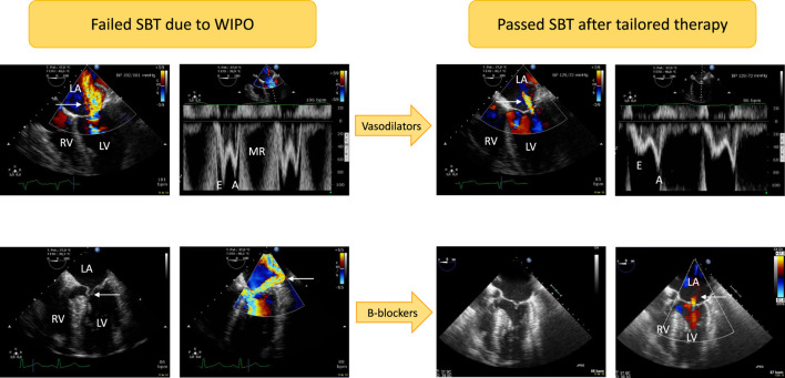
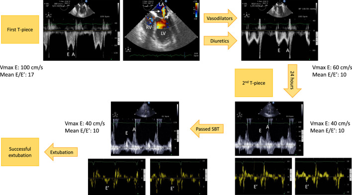
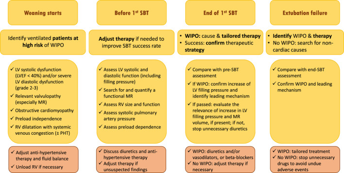
Weaning a critically-ill patient from the ventilator is a crucial step in global management. This manuscript details physiological changes induced by altered heart-lung interactions during the weaning process, illustrates the main mechanisms which could lead to weaning failure of cardiac origin, and discuss a tailored management based on the monitoring of changes in central hemodynamics during weaning. The transition from positive-pressure ventilation to spontaneous breathing results in abrupt hemodynamic and metabolic changes secondary to rapidly modified heart-lung interactions, sudden changes in cardiac loading conditions, and increased oxygen demand. These modifications may elicit an excessive burden on both the respiratory and cardiovascular systems, result in a rapid and marked increase of left ventricular filling pressure, and ultimately result in a weaning-induced pulmonary oedema (WIPO). The T-piece trial induces the greatest burden on respiratory and cardiocirculatory function when compared to spontaneous breathing trial using pressure support ventilation with positive or zero end-expiratory pressure. Since LV overload is the mainstay of WIPO, positive fluid balance and SBT-induced acute hypertension are the most frequently reported mechanisms of weaning failure of cardiac origin. Although the diagnosis of WIPO historically relied on an abrupt elevation of pulmonary artery occlusion pressure measured during right heart catheterization, it is nowadays commonly documented by echocardiography Doppler. This non-invasive approach is best suited for identifying high-risk patients, depicting the origin of WIPO, and tailoring individual management. Whether this strategy increases the success rate of weaning needs to be evaluated in a population at high risk of weaning failure of cardiac origin.

Keywords: echocardiography; heart-lung interactions; mechanical ventilation; pulmonary edema; weaning.


Conflict of interest statement

The author declares that the research was conducted in the absence of any commercial or financial relationships that could be construed as a potential conflict of interest.

Figures

FIGURE 1
FIGURE 1
Illustrative cases of acute severe mitral regurgitation induced by abrupt changes in left ventricular loading conditions and increased adrenergic tone during a T-piece trial. Patients developed severe weaning-induced pulmonary edema and were promptly reconnected to the ventilator to be assessed using transesophageal echocardiography. In the first patient (upper panels) with acute hypertension (192/101 mmHg), a massive functional mitral regurgitation (central jet on color Doppler mapping, upper left panel, arrow) with markedly elevated mitral Doppler velocities (upper left middle panel; E wave maximal velocity: 1 m/s) was evidenced. Nitrates were administered intravenously as sequential boli to rapidly normalize blood pressure (129/72 mmHg). This allowed to dramatically reduce the volume of mitral regurgitation as reflected by a marked decrease of color Doppler jet area (upper right middle panel, arrow) as well as mitral Doppler velocities (upper right panel; maximal E wave velocity: 0.5 m/s). In the second patient (lower panels), echocardiography depicted a dynamic obstruction of left ventricular outflow tract by an anterior motion of the mitral valve (lower left panel, arrow). This resulted in high end-systolic pressure gradient and associated massive eccentric mitral regurgitation (lower middle left panel, arrow). Beta-blockers were successfully used: both the dynamic left ventricular outflow tract obstruction and mitral regurgitation fully resolved as depicted by two-dimensional imaging (lower middle right panel; end-systole) and color Doppler mapping which only disclosed a residual trivial central mitral regurgitation (lower right panel, arrow). Abbreviations: LA, left atrium; LV, left ventricle; RV, right ventricle; MR: mitral regurgitation.
FIGURE 2
FIGURE 2
Example of hemodynamic monitoring using echocardiography in a patient with left ventricular systolic dysfunction secondary to an ischemic heart disease who developed a weaning-induced pulmonary edema during the first T-piece trial which induced high blood pressure. Immediately after having reconnected the patient to the ventilator, a transesophageal echocardiography was performed. Mitral Doppler depicted elevated E wave velocity and mean E/E′ ratio, consistent with increased left ventricular filling pressure (upper left panel). There was only a mild functional mitral regurgitation (upper middle panel, arrow). Treatment associated short half-life sedative, ACE inhibitors and diuretics. With normalization of blood pressure and fluid removal, the mitral Doppler profile remained unchanged (E/A ratio ≈1), but velocities significantly decreased as well as mean E/E′ ratio (upper right panel). This allowed to perform a second T-piece trial 24 h later. Immediately before the procedure, mitral E wave velocity remained low, E/A ratio was <1, and mean E/E′ remained stable, denoting the absence of new rise in left ventricular filling pressure (3 lower right panels). The second spontaneous breathing trial was clinically successful. Echocardiography at the end of the 30-min T-piece trial depicted similar mitral Doppler velocity values, hence left ventricular filling pressure (3 lower left panels). The patient was then extubated successfully without any change of ongoing treatment. Abbreviations: LA, left atrium; LV, left ventricle; RV, right ventricle.
FIGURE 3
FIGURE 3
Proposed strategy of use of echocardiography monitoring in ventilated patients who are ready to be weaned from the ventilator and potentially at risk of weaning failure of cardiac origin (see text for details). Abbreviations: SBT, spontaneous breathing trial; WIPO, weaning-induced pulmonary edema; LV, left ventricle; EF, ejection fraction; MR, mitral regurgitation; RV, right ventricle; PHT, pulmonary hypertension.

Similar articles

Cited by

References

    1. Adamopoulos C., Tsagourias M., Arvaniti K., Veroniki F., Matamis D. (2005). Weaning failure from mechanical ventilation due to hypertrophic obstructive cardiomyopathy. Intensive Care Med. 31, 734–737. 10.1007/s00134-005-2616-2 - DOI - PubMed
    1. Ait-Oufella H., Tharaux P. L., Baudel J. L., Vandermeersch S., Meyer P., Tonnellier M., et al. (2007). Variation in natriuretic peptides and mitral flow indexes during successful ventilatory weaning: A preliminary study. Intensive Care Med. 33, 1183–1186. 10.1007/s00134-007-0627-x - DOI - PubMed
    1. Bataille B., Riu B., Moussot P. E., Mari A., Brunel E., (2014). Integrated use of bedside lung ultrasound and echocardiography in acute respiratory failure: A prospective observational study in ICU. Chest 146, 1586–1593. 10.1378/chest.14-0681 - DOI - PubMed
    1. Bedet A., Tomberli F., Prat G., Bailly P., Kouatchet A., Mortaza S., et al. (2019). Myocardial ischemia during ventilator weaning: A prospective multicenter cohort study. Crit. Care 23, 321. 10.1186/s13054-019-2601-8 - DOI - PMC - PubMed
    1. Beduneau G., Pham T., Schortgen F., Piquilloud L., Zogheib E., Jonas M., et al. (2017). Epidemiology of weaning outcome according to a new definition. The WIND study. Am. J. Resp. Crit. Care Med. 195, 772–783. 10.1164/rccm.201602-0320OC - DOI - PubMed

LinkOut - more resources