. 2023 Sep 8:14:1259567.
doi: 10.3389/fphys.2023.1259567. eCollection 2023.

The accordion technique enhances bone regeneration via angiogenesis factor in a rat distraction osteogenesis model

Affiliations

The accordion technique enhances bone regeneration via angiogenesis factor in a rat distraction osteogenesis model

Kai Liu et al. Front Physiol. .

Abstract

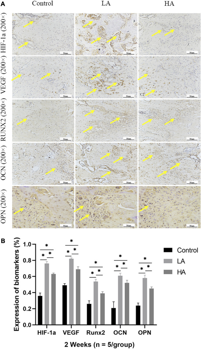
Objective: The purpose of this study was to observe the effect of the accordion technique (AT) during the distraction phase on chondrogenesis and bone regeneration in a rat femoral distraction osteogenesis (DO) model, and investigate its potential mechanism for reducing the total treatment time of DO. Methods: Fifty-four male Sprague-Dawley (SD) rats that were specific-pathogen-free (SPF) were subjected to DO surgery on the right femur. The distraction rate was 0.5 mm/day for 10 days, following a latency period of 5 days. Rats were randomly divided into Control (no AT, n = 18), Group LA (low amplitude with AT, n = 18), and Group HA (high amplitude with AT, n = 18) according to different AT protocols in the distraction phase. Rats were respectively euthanized by anesthesia overdose at 2, 4 and 6 weeks of the consolidation phase, and the femurs were harvested. Digital radiography, micro-computed tomography (micro-CT), biomechanical tests, and histomorphological analysis were used to assess the quality of regenerated bone in the distraction area. Results: Digital radiographic, micro-CT, biomechanical tests, and histological analysis revealed an increase in early-stage callus formation (p < 0.05) and improved blood supply to the callus tissue in Group LA, as compared to both the Control and Group HA. The enhanced differentiation of fibrous and cartilaginous tissue into bone tissue was also observed in Group LA, leading to improved strength and stiffness (p < 0.05) of the regenerated bone at 6 weeks of the consolidation phase. The angiogenic (hypoxia-inducible factor-1α (HIF-1α) and vascular endothelial growth factor (VEGF), p < 0.05) and osteogenic (runt-related transcription factor 2 (RUNX2), osteocalcin (OCN) and osteopontin (OPN), p < 0.05) biomarkers were higher expressed in Group LA at 2 and 4 weeks of consolidation phase, whereas decreased at 6 weeks of consolidation phase. Conclusion: The application of AT with low amplitude during the distraction phase can enhance chondrogenesis and bone regeneration by activating the angiogenesis factor pathway and upregulating the expression of osteogenic-related biomarkers such as HIF-1α, VEGF, RUNX2, OCN, and OPN.

Keywords: accordion maneuver; angiogenesis factor; bone regeneration; bone remodeling; distraction osteogenesis.


Conflict of interest statement

The authors declare that the research was conducted in the absence of any commercial or financial relationships that could be construed as a potential conflict of interest.

Figures

FIGURE 1
FIGURE 1
The general images and X-ray images of ROI until the 6-week consolidation phase terminated. (A) X-ray of the distraction area, the regenerated bone of the distraction area in Group LA (red areas) was markedly increased (yellow arrows) at the consolidation phase. (B) The general appearance of the distraction area (red areas) showed that recanalization of the medullary cavity (yellow arrows) was received in Group LA at 6 weeks of consolidation.
FIGURE 2
FIGURE 2
Micro-CT evaluation of ROI at 6 weeks of consolidation phase. (A) Representative 3D micro-CT images of the distraction area (red areas) at 6 weeks of the consolidation phase showed that recanalization of the medullary cavity (yellow arrows) was received in Group LA. However, narrow gaps in the distraction area were observed in the Control and Group HA. (B–H) Quantitative evaluation of the values of BMD, BV, TV, BV/TV, Tb. N, Tb. Th, Tb. Th, and Tb. Sp in Group LA and Group HA were significantly higher than the Control (*p < 0.05; ns, p > 0.05).
FIGURE 3
FIGURE 3
Results of mechanical values of ROI after 6 weeks of consolidation. (A–C) The non-normalized stiffness (N/mm), ultimate load (N), and energy to failure (N/mm2) in Group LA and Group HA were significantly better than those in Control (*p < 0.05).
FIGURE 4
FIGURE 4
The histomorphological analysis of ROI (low-magnification × 50, and high-magnification × 200) during the consolidation phase. (A) H&E staining revealed more microvessels, bone trabeculae, and callus formation (yellow arrows) in Group LA compared to the other two groups at 2 and 4 weeks of the consolidation phase. The two osteogenic fronts (red arrows) originated at the proximal and distal ends of the distraction area and gradually progressed towards the center until the medullary cavity was completely recanalized. By 6 weeks, the recanalization of the medullary cavity (black arrows) in the distraction area of Group LA was achieved. In contrast, both the Control and Group HA still exhibited a significant amount of callus and immature cartilage-like tissue (yellow arrows), with no recanalization of the medullary cavity. (B) Safranin O-Fast Green staining demonstrated increased chondrogenesis (red staining indicated by yellow arrows, primarily composed of prechondrocytes) and bone formation (green staining) in Group LA at 2 and 4 weeks of the consolidation phase. (C) Masson trichrome staining indicated enhanced chondrogenesis (blue collagen fibers and cartilage, yellow arrows) and bone regeneration in Group LA at 2 and 4 weeks of the consolidation phase. Moreover, these cartilage-like tissues gradually transformed into mature bone tissue with medullary recanalization (black arrows) by 6 weeks of the consolidation phase.
FIGURE 5
FIGURE 5
Immunohistochemistry images (high-magnification 200 ×) in the three groups at 2 weeks of consolidation phase (A) In Group LA, the positive staining area or cells (yellow arrows) were predominantly localized in the central region of the distraction area, whereas in Group HA and the Control group, they were mainly observed at the edge of the distraction area. (B) At the 2 weeks of consolidation phase, the semi-quantitative measurements demonstrated a significant upregulation of HIF-1α, VEGF, RUNX2, OCN, and OPN expression in Group LA, compared to Group HA and the Control (*p < 0.05).
FIGURE 6
FIGURE 6
Immunohistochemistry images (high-magnification 200 ×) in the three groups at 4 weeks of consolidation phase (A) At the 4 weeks of consolidation phase, as osteoblasts and chondroblasts differentiate into mature osteocytes and mineralization progresses, the expression of angiogenic (HIF-1α, VEGF) and osteogenesis-related proteins (RUNX2, OCN, and OPN) progressively decreased in all three groups. Notably, in Group LA, the positive staining area or cells (yellow arrows) remained predominantly concentrated in the central region of the distraction area. However, in Group HA and the Control group, they were primarily observed at the edge of the distraction area. (B) The semi-quantitative measurements showed that the above biomarkers were highly expressed in Group LA, compared to Group HA and the Control (*p < 0.05).
FIGURE 7
FIGURE 7
Immunohistochemistry images (high-magnification 200 ×) in the three groups at 6 weeks of consolidation phase (A) At the 6 weeks of consolidation phase, the positive staining area or cells (yellow arrows) in the Control were primarily concentrated in the central region of the distraction area. However, there were only a few positively stained areas or cells in Group LA and Group HA, which can be attributed to the recanalization of the medullary cavity and bone remodeling. (B) The semi-quantitative measurements revealed that the expression levels of HIF-1α, VEGF, RUNX2, OCN, and OPN were higher in the Control group compared to both Group LA and Group HA (*p < 0.05).

Similar articles

Cited by

References

    1. Ai-Aql Z. S., Alagl A. S., Graves D. T., Gerstenfeld L. C., Einhorn T. A. (2008). Molecular mechanisms controlling bone formation during fracture healing and distraction osteogenesis. J. Dent. Res. 87 (2), 107–118. 10.1177/154405910808700215 - DOI - PMC - PubMed
    1. Alzahrani M. M., Anam E., AlQahtani S. M., Makhdom A. M., Hamdy R. C. (2018). Strategies of enhancing bone regenerate formation in distraction osteogenesis. Connect. Tissue Res. 59 (1), 1–11. 10.1080/03008207.2017.1288725 - DOI - PubMed
    1. Aronson J. (1994). Temporal and spatial increases in blood flow during distraction osteogenesis. Clin. Orthop. Relat. Res. 301, 124–131. 10.1097/00003086-199404000-00020 - DOI - PubMed
    1. Bezstarosti H., Metsemakers W. J., van Lieshout E., Voskamp L. W., Kortram K., McNally M. A., et al. (2021). Management of critical-sized bone defects in the treatment of fracture-related infection: A systematic review and pooled analysis. Arch. Orthop. Trauma Surg. 141 (7), 1215–1230. 10.1007/s00402-020-03525-0 - DOI - PMC - PubMed
    1. Borzunov D. Y. (2012). Long bone reconstruction using multilevel lengthening of bone defect fragments. Int. Orthop. 36 (8), 1695–1700. 10.1007/s00264-012-1562-1 - DOI - PMC - PubMed