The spleen as a possible source of serine protease inhibitors and migrating monocytes required for liver regeneration after 70% resection in mice
- PMID: 37745290
- PMCID: PMC10512715
- DOI: 10.3389/fcell.2023.1241819
The spleen as a possible source of serine protease inhibitors and migrating monocytes required for liver regeneration after 70% resection in mice
Abstract
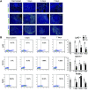
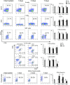
Introduction: The role of the immune system in liver repair is fundamentally complex and most likely involves the spleen. The close connection between the two organs via the portal vein enables delivery of splenic cytokines and living cells to the liver. This study evaluates expression of inflammation-related genes and assesses the dynamics of monocyte-macrophage and lymphocyte populations of the spleen during the recovery from 70% hepatectomy in mice. Methods: The study used the established mouse model of 70% liver volume resection. The animals were sacrificed 24 h, 72 h or 7 days post-intervention and splenic tissues were collected for analysis: Clariom™ S transcriptomic assay, immunohistochemistry for proliferation marker Ki-67 and macrophage markers, and flow cytometry for lymphocyte and macrophage markers. Results: The loss and regeneration of 70% liver volume affected the cytological architecture and gene expression profiles of the spleen. The tests revealed significant reduction in cell counts for Ki-67+ cells and CD115+ macrophages on day 1, Ly6C + cells on days 1, 3 and 7, and CD3+CD8+ cytotoxic lymphocytes on day 7. The transcriptomic analysis revealed significant activation of protease inhibitor genes Serpina3n, Stfa2 and Stfa2l1 and decreased expression of cell cycle regulatory genes on day 1, mirrored by inverse dynamics observed on day 7. Discussion and conclusion: Splenic homeostasis is significantly affected by massive loss in liver volume. High levels of protease inhibitors indicated by increased expression of corresponding genes on day 1 may play an anti-inflammatory role upon reaching the regenerating liver via the portal vein. Leukocyte populations of the spleen react by a slow-down in proliferation. A transient decrease in the local CD115+ and Ly6C+ cell counts may indicate migration of splenic monocytes-macrophages to the liver.
Keywords: liver; lymphocytes; macrophages; regeneration; spleen.
Copyright © 2023 Elchaninov, Vishnyakova, Kuznetsova, Gantsova, Kiseleva, Lokhonina, Antonova, Mamedov, Soboleva, Trofimov, Fatkhudinov and Sukhikh.
Conflict of interest statement
The authors declare that the research was conducted in the absence of any commercial or financial relationships that could be construed as a potential conflict of interest.
Figures


Similar articles
-
Cellular effects of splenectomy on liver regeneration after 70% resection.Front Cell Dev Biol. 2025 May 1;13:1561815. doi: 10.3389/fcell.2025.1561815. eCollection 2025. Front Cell Dev Biol. 2025. PMID: 40376613 Free PMC article.
-
Macro- and microtranscriptomic evidence of the monocyte recruitment to regenerating liver after partial hepatectomy in mouse model.Biomed Pharmacother. 2021 Jun;138:111516. doi: 10.1016/j.biopha.2021.111516. Epub 2021 Mar 23. Biomed Pharmacother. 2021. PMID: 33765583
-
Spleen regeneration after subcutaneous heterotopic autotransplantation in a mouse model.Biol Res. 2023 Mar 29;56(1):15. doi: 10.1186/s40659-023-00427-4. Biol Res. 2023. PMID: 36991509 Free PMC article.
-
Hypergravity-induced immunomodulation in a rodent model: lymphocytes and lymphoid organs.J Gravit Physiol. 2002 Dec;9(2):15-27. J Gravit Physiol. 2002. PMID: 14638456
-
CD11b + CD43 hi Ly6C lo splenocyte-derived macrophages exacerbate liver fibrosis via spleen-liver axis.Hepatology. 2023 May 1;77(5):1612-1629. doi: 10.1002/hep.32782. Epub 2023 Apr 17. Hepatology. 2023. PMID: 36098707 Free PMC article.
Cited by
-
Cellular effects of splenectomy on liver regeneration after 70% resection.Front Cell Dev Biol. 2025 May 1;13:1561815. doi: 10.3389/fcell.2025.1561815. eCollection 2025. Front Cell Dev Biol. 2025. PMID: 40376613 Free PMC article.
-
Computational inference of chemokine-mediated roles for the vagus nerve in modulating intra- and inter-tissue inflammation.Front Syst Biol. 2024 Feb 15;4:1266279. doi: 10.3389/fsysb.2024.1266279. eCollection 2024. Front Syst Biol. 2024. PMID: 40809119 Free PMC article.
References
-
- Babaeva A. G. (1989). Cellular and humoral immunity factors as regulators of regenerative morphogenesis. Ontogenez 20, 453–460. Available at: http://www.ncbi.nlm.nih.gov/pubmed/2531358 (Accessed November 11, 2019). - PubMed
-
- Babaeva A. G., Zotikov E. A. (1987). Immunology of processes of adaptive growth, proliferation and their disorders. Moscow: Medicine.
LinkOut - more resources
Full Text Sources
Molecular Biology Databases
Research Materials
Miscellaneous

