[Preprint]. 2023 Sep 13:2023.09.11.557226.
doi: 10.1101/2023.09.11.557226.

Clinicopathologic Dissociation: Robust Lafora Body Accumulation in Malin KO Mice Without Observable Changes in Home-cage Behavior

Affiliations

Clinicopathologic Dissociation: Robust Lafora Body Accumulation in Malin KO Mice Without Observable Changes in Home-cage Behavior

Vaishnav Krishnan et al. bioRxiv. .

Update in

Abstract

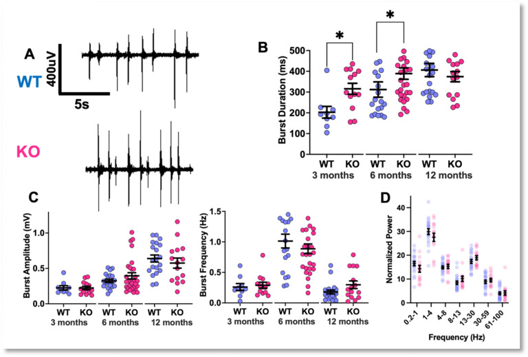
Lafora Disease (LD) is a syndrome of progressive myoclonic epilepsy and cumulative neurocognitive deterioration caused by recessively inherited genetic lesions of EPM2A (laforin) or NHLRC1 (malin). Neuropsychiatric symptomatology in LD is thought to be directly downstream of neuronal and astrocytic polyglucosan aggregates, termed Lafora bodies (LBs), which faithfully accumulate in an age-dependent manner in all mouse models of LD. In this study, we applied home-cage monitoring to examine the extent of neurobehavioral deterioration in a model of malin-deficient LD, as a means to identify robust preclinical endpoints that may guide the selection of novel genetic treatments. At 6 weeks, ~6-7 months and ~12 months of age, malin deficient mice ("KO") and wild type (WT) littermates underwent a standardized home-cage behavioral assessment designed to non-obtrusively appraise features of rest/arousal, consumptive behaviors, risk aversion and voluntary wheel-running. At all timepoints, and over a range of metrics that we report transparently, WT and KO mice were essentially indistinguishable. In contrast, within WT mice compared across timepoints, we identified age-related nocturnal hypoactivity, diminished sucrose preference and reduced wheel-running. Neuropathological examinations in subsets of the same mice revealed expected age dependent LB accumulation, gliosis and microglial activation in cortical and subcortical brain regions. At 12 months of age, despite the burden of neocortical LBs, we did not identify spontaneous seizures during an electroencephalographic (EEG) survey, and KO and WT mice exhibited similar spectral EEG features. Using an in vitro assay of neocortical function, paroxysmal increases in network activity (UP states) in KO slices were more prolonged at 3 and 6 months of age, but were similar to WT at 12 months. KO mice displayed a distinct response to pentylenetetrazole, with a greater incidence of clonic seizures and a more pronounced post-ictal suppression of movement, feeding and drinking behavior. Together, these results highlight a stark clinicopathologic dissociation in a mouse model of LD, where LBs accrue substantially without clinically meaningful changes in overall wellbeing. Our findings allude to a delay between LB accumulation and neurobehavioral decline: one that may provide a window for treatment, and whose precise duration may be difficult to ascertain within the typical lifespan of a laboratory mouse.

Keywords: Lafora body disease; astrogliosis; glycogen storage; home-cage behavior; malin; polyglucosan.


Conflict of interest statement

Conflict of interest disclosure: The authors have no relevant conflicting interests to disclose.

Figures

Figure 1.
Figure 1.. Lafora body (LB) Accumulation and Initial Home-Cage Response.
A: Representative PAS-D images showing LB accumulation (TOP) and immunohistochemical assessments of LB accumulation (GS1), astrogliosis (GFAP) and microglial activation (Iba1). Scale bar: 50 μm. B: Home-cage structure and representative aerial video snapshot. C: Raster plot of distances traversed every minute of the 2-hour long introduction trial for every mouse, with measures of licking, shelter and feeder engagement. D: Heatmaps (left) and trackmaps (right) for a representative WT and KO mouse. Mean ± s.e.m shown for all.
Figure 2.
Figure 2.. Baseline Recordings.
A: Hourly horizontal distances within the home-cage (lights OFF between 1700 and 0500), total distances, and ultradian rhythms of activity (Lomb-Scargle Periodogram). BOTTOM: Raster plot of distances moved every minute of the day, displaying active/inactive state structure. B: KO mice displayed similar bout structure and timing of “sleep” (measured noninvasively). C: Averaged time budgets for WT and KO mice. D: WT and KO mice displayed similar sucrose preference and lick macrostructure. E: WT and KO mice displayed similar feeding entries and durations. Mean ± s.e.m shown for all. See Fig.1A for sample sizes.
Figure 3.
Figure 3.. Home-cage provocative maneuvers.
A: Changes to horizontal activity and sheltering in response to a single hour of light-spot stimulation. B: Distances and sheltering responses to a single 60s-long beep stimulus (2300hz tone). C: Wheel rotations accumulated during a 23h-long wheel-running trial. D: Distance/sheltering behavior following cage-swap. Mean ± s.e.m shown for all. See Fig.1 for sample sizes.
Figure 4.
Figure 4.. Changes in Home-cage Behavior with Age.
A: On baseline day 2, compared with 6-week old mice, older WT cohorts displayed diminished sucrose preference (F2,56 = 2.94, p = 0.06) and feeding durations (F2,56 = 10.55, p<0,0001). B: Measures of sleep timing and duration were unchanged. C,D: 6-month and 1-year old mice displayed a blunted response to light-spot and BEEP stimulation. E: Older mice displayed fewer wheel rotations (F2,56 = 9.10, p<0.001). Mean ± s.e.m shown for all. *, **, ***, **** depict p<0.05, <0.01, <0.001 or <0.0001 respectively.
Figure 5.
Figure 5.. EEG and PTZ Responses.
A: Representative single-channel electrocorticography from 1-year old WT and KO mice. B: EEG power spectra calculated during wakefulness. C: Representative EEG responses to a single intraperitoneal injection of PTZ (60mg/kg), demonstrating a prolonged epoch of spike/wave discharges, followed by a discrete epoch of evolving rhythmicity. Red bars annotate epochs of absent EEG signal while the mouse receives the intraperitoneal injection. D: Distance and sheltering responses to a single subconvulsant PTZ injection (30mg/kg), with a tally of convulsive events (inset) for both WT (n=14) and KO (n = 18). E: Post-ictal period home-cage metrics, revealing a comparative increase in sheltering and reduction in feeding in KO mice. Mean ± s.e.m shown for all. * depicts p<0.05.
Figure 6:
Figure 6:. UP States in WT vs KO Somatosensory Cortex.
A: Example traces of extracellular recordings in brain slices exhibiting spontaneously occurring activity bursts (from 12-month-old mice). B: The average duration of activity bursts in MKO slices is longer at 3 and 6 months of age, but not at 12 months (Mann-Whitney test, *p< 0.05) C: Burst amplitudes and frequencies. D: Relative power over all activity. Mean ± s.e.m shown for all. Sample sizes at 3 months: WT (9 slices, 4 mice), KO (13 slices, 5 mice). 6 months: WT (19 slices, 6 mice), KO (29 slices, 7 mice). 12 months: WT (12 slices, 6 mice), KO (15 slices, 5 mice).
Figure 7:
Figure 7:
Lafora body accumulation in mouse piriform cortex (malin KO, 12 months of age).

References

    1. Mitra S, Gumusgoz E, Minassian BA. Lafora disease: Current biology and therapeutic approaches. Rev Neurol (Paris). 2022;178(4):315–325. - PMC - PubMed
    1. Nitschke F, Ahonen SJ, Nitschke S, Mitra S, Minassian BA. Lafora disease - from pathogenesis to treatment strategies. Nat Rev Neurol. 2018;14(10):606–617. - PMC - PubMed
    1. Brenner D, Baumgartner T, von Spiczak S, et al. Genotypes and phenotypes of patients with Lafora disease living in Germany. Neurol Res Pract. 2019;1. - PMC - PubMed
    1. Riva A, Orsini A, Scala M, et al. Italian cohort of Lafora disease: Clinical features, disease evolution, and genotype-phenotype correlations. J Neurol Sci. 2021;424:117409. - PMC - PubMed
    1. Singh S, Satishchandra P, Shankar SK, Ganesh S. Lafora disease in the Indian population: EPM2A and NHLRC1 gene mutations and their impact on subcellular localization of laforin and malin. Hum Mutat. 2008;29(6):E1–12. - PubMed

Publication types