[Preprint]. 2023 Sep 17:2023.09.14.557681.
doi: 10.1101/2023.09.14.557681.

A latent cardiomyocyte regeneration potential in human heart disease

Affiliations

A latent cardiomyocyte regeneration potential in human heart disease

Wouter Derks et al. bioRxiv. .

Update in

  • A Latent Cardiomyocyte Regeneration Potential in Human Heart Disease.
    Derks W, Rode J, Collin S, Rost F, Heinke P, Hariharan A, Pickel L, Simonova I, Lázár E, Graham E, Jashari R, Andrä M, Jeppsson A, Salehpour M, Alkass K, Druid H, Kyriakopoulos CP, Taleb I, Shankar TS, Selzman CH, Sadek H, Jovinge S, Brusch L, Frisén J, Drakos S, Bergmann O. Derks W, et al. Circulation. 2025 Jan 21;151(3):245-256. doi: 10.1161/CIRCULATIONAHA.123.067156. Epub 2024 Nov 21. Circulation. 2025. PMID: 39569515 Free PMC article.

Abstract

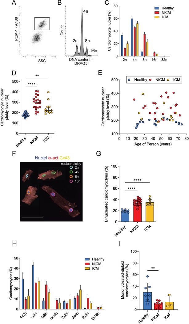
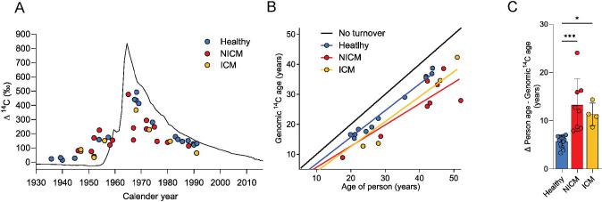
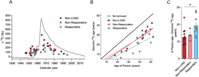
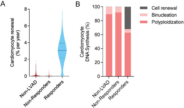
Cardiomyocytes in the adult human heart show a regenerative capacity, with an annual renewal rate around 0.5%. Whether this regenerative capacity of human cardiomyocytes is employed in heart failure has been controversial. Using retrospective 14C birth dating we analyzed cardiomyocyte renewal in patients with end-stage heart failure. We show that cardiomyocyte generation is minimal in end-stage heart failure patients at rates 18-50 times lower compared to the healthy heart. However, patients receiving left ventricle support device therapy, who showed significant functional and structural cardiac improvement, had a >6-fold increase in cardiomyocyte renewal relative to the healthy heart. Our findings reveal a substantial cardiomyocyte regeneration potential in human heart disease, which could be exploited therapeutically.


Conflict of interest statement

The authors declare no competing interests.

Figures

Figure 1.
Figure 1.. Isolation of human cardiomyocytes and ploidy assessment
(A) Flow cytometry-based sorting enables identification and isolation of cardiomyocyte nuclei with antibodies against PCM-1 (inlet). (B) Flow cytometry analysis of cardiomyocyte nuclei DNA content reveals their ploidy profile (2n = diploid, 4n = tetraploid, 8n = octaploid, 16n = hexadecaploid; shown is a representative NICM sample). (C) The distribution of cardiomyocyte nuclear ploidy populations shows a shift to higher ploidy levels in NICM (n=21) and ICM patients (n=11) compared to healthy adults (n=11, from (2)). (D) Cardiomyocyte nuclear ploidy level determined by flow cytometry is higher in both NICM (n=21) and ICM patients (n=11) than in healthy adults (n=11) (e.g. 100% corresponds to only diploid nuclei; 200% to only tetraploid nuclei) (Kruskal-Wallis one-way ANOVA on Ranks, H=30.3, p < 0.001, post hoc Dunn’s ** p=0.0063, **** p<0.0001), lines show median with interquartile range. (E) Ploidy increase in cardiomyopathy is not related to the age of the adult patient (NICM R=0.26, p=0.26; ICM R=0.49, p=0.11). (F) Image cytometry identifies cardiomyocyte ploidy classes. Cardiomyocytes were digested from tissue samples and stained for α-actinin (α-act) and connexin-43 (Cx43). The integrated intensity of the DNA dye was used to designate the nuclear ploidy class of each imaged nucleus, as shown by the overlay in different colors on the image. Cx43 enabled to determine if cardiomyocytes were intactly dissociated at the intercalated discs. Figure shows a compilation of several cardiomyocytes of different ploidy and nucleation levels. Scale bar size is 100 μm (G) The percentage of binucleated cardiomyocytes determined by image cytometry is higher in NICM (n=27) and ICM hearts (n=8) compared to healthy (n=8) (Ordinary one-way ANOVA, F=32.1; p < 0.0001, post hoc Tukey, **** p<0.0001). (H) Both binucleation and nuclear ploidy levels were determined in isolated cardiomyocytes from healthy (n=7) and pathological hearts (NICM n=8, ICM n=3) with image cytometry. (I) The percentage of mononucleated diploid cardiomyocytes is smaller in NICM 9.7% ± 5.2% and ICM 13.13% ± 10.56% compared to healthy hearts 29.7% ± 15.8% (Kruskal-Wallis one-way-ANOVA on ranks; H = 7.7; p = 0.021, ** p=0.0073).
Figure 2.
Figure 2.. 14C levels indicate increased DNA synthesis in cardiomyocytes of failing hearts
(A) Presentation of 14C data. The black curve indicates the historic atmospheric 14C concentrations. 14C measurements from heart samples are plotted as colored dots on the date of subject birth. Genomic 14C values of cardiomyocytes in healthy hearts (blue dots, n=18, data taken from Bergmann et al., 2015) are close to atmospheric values at the time of birth, indicating a limited postnatal and adult renewal of cells. The deviation of 14C values of cardiomyocytes in NICM (red dots, n=16) and ICM hearts (orange dots, n=8) from the atmospheric 14C curve suggest genomic DNA turnover. (B) The estimated genomic 14C age of cardiomyocytes was calculated from subjects with post-bomb birth dates and plotted at the person’s age. The black line indicates no turnover. (C) Both NICM (n=8) and ICM samples (n=4) show a higher deviation of the estimated genomic 14C age from the subject’s age compared to a healthy heart (n=12), suggesting increased DNA synthesis (Ordinary one-way ANOVA, F=12.38; p < 0.0002, post hoc Dunnet’s multiple comparison, *** p=0.0002, *p=0.017).
Figure 3
Figure 3. Mathematical modeling the renewal dynamics of cardiomyocytes in failing hearts
(A) With the two-phase renewal model the annual cardiomyocyte renewal rate can be determined before and after disease onset. In the disease phase, the percentage of newly formed cardiomyocytes drops from 0.55% [0.46%;0.64%] (median and interquartile range) to 0.03% [0.002%;0.45%] (median and interquartile range) in NICM, and 0.01% [0.001%;0.13%] (median and interquartile range) in ICM. (B) The annual amount of DNA synthesis modelled shows an increase in both NICM and ICM compared to healthy. (C) Percentage of DNA synthesis attributed to cell renewal, binucleation and nuclear polyploidization estimated in diseased hearts. After disease onset in NICM and ICM patients only < 0.3% of DNA synthesis can be attributed to proliferation-based cell renewal.
Figure 4.
Figure 4.. 14C levels in cardiomyocytes of LVAD-supported hearts
(A) Presentation of 14C data from unloaded hearts. 14C measurements from heart samples are plotted as colored dots on the date of subject birth for diseased non-LVAD hearts (red dots, n=24, these correspond to NICM and ICM samples as shown in figure 2A), LVAD non-responders (n=13) and LVAD responders (n=15). The location of the dots from responder subjects suggests an increased deviation from the atmospheric 14C curve compared to non-LVAD subjects. (B) The estimated genomic 14C age of cardiomyocytes was calculated from subjects with post-bomb birth dates and plotted at the person’s age. The black line indicates no turnover. (C) Responders (n=8) show genomic 14C age which is on average 19.25 years younger than the person, compared to 13.94 years for LVAD non-responders (n=3) and 12.61 years for non-LVAD patients (n=12).
Figure 5.
Figure 5.. Mechanical unloading increases cardiomyocyte renewal in LVAD responders
(A) Cardiomyocyte renewal rate as determined using the two-phase renewal model. In cardiomyopathy, the percentage of newly formed cardiomyocytes per year is 3.07% [2.16%; 3.94] in LVAD responders compared to 0.03% in the non-LVAD group [0.002%;0.35%] and 0.02% in LVAD non-responders [0.001%;0.20%] (median and interquartile range). (B) Percentage of DNA synthesis attributed to cell renewal, binucleation and nuclear polyploidization determined at time of sample collection. In LVAD responders, 32.0% of all DNA synthesis can be attributed to proliferation-based cell renewal, in contrast to only 0.26% and 0.33% in non-responders and non-LVAD patients.

References

    1. Nakamura K., Murry C. E., Function Follows Form- A Review of Cardiac Cell Therapy. Circ J 83, 2399–2412 (2019). - PMC - PubMed
    1. Bergmann O. et al., Dynamics of Cell Generation and Turnover in the Human Heart. Cell 161, 1566–1575 (2015). - PubMed
    1. Bergmann O. et al., Evidence for cardiomyocyte renewal in humans. Science 324, 98–102 (2009). - PMC - PubMed
    1. Chien K. R. et al., Regenerating the field of cardiovascular cell therapy. Nat Biotechnol 37, 232–237 (2019). - PubMed
    1. Eschenhagen T. et al., Cardiomyocyte Regeneration: A Consensus Statement. Circulation 136, 680–686 (2017). - PMC - PubMed

Publication types