[Preprint]. 2023 Sep 14:2023.09.11.557277.
doi: 10.1101/2023.09.11.557277.

Interferon lambda restricts herpes simplex virus skin disease by suppressing neutrophil-mediated pathology

Affiliations

Interferon lambda restricts herpes simplex virus skin disease by suppressing neutrophil-mediated pathology

Drake T Philip et al. bioRxiv. .

Update in

Abstract

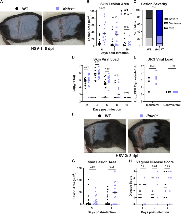
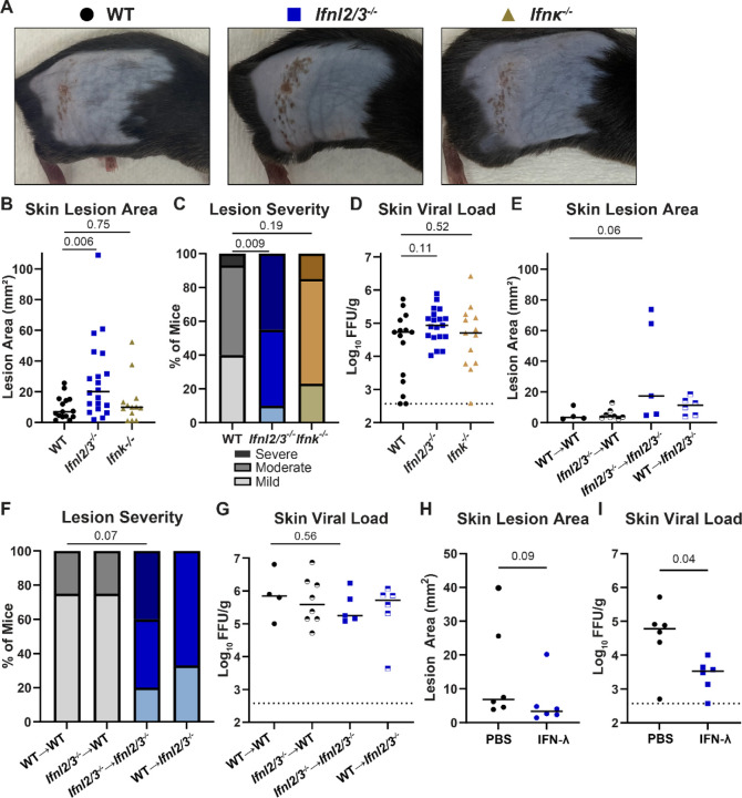
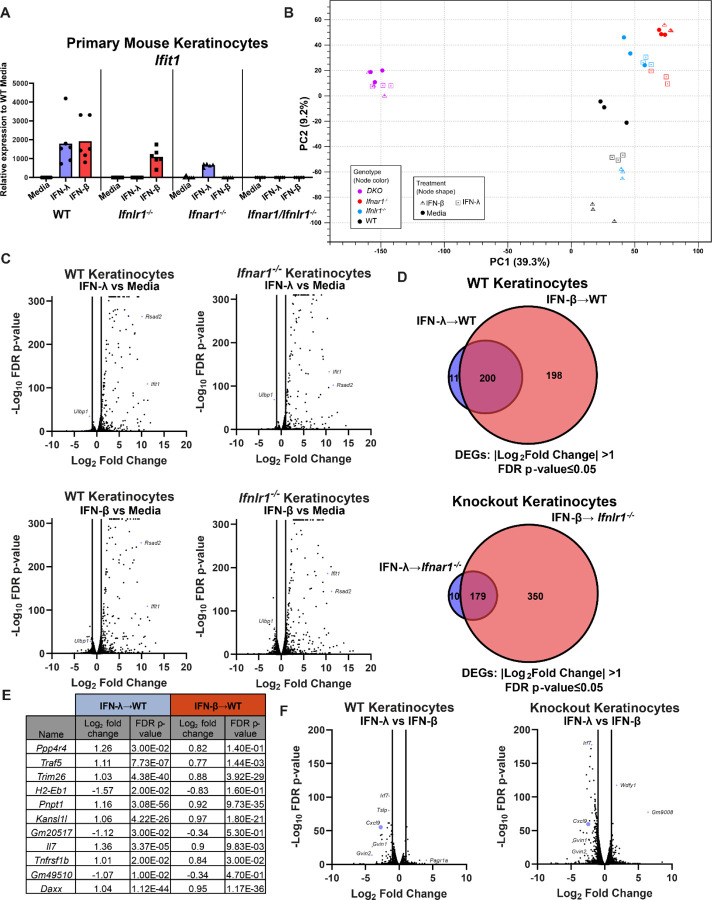
Type III interferons (IFN-λ) are antiviral and immunomodulatory cytokines that have been best characterized in respiratory and gastrointestinal infections, but the effects of IFN-λ against skin infections have not been extensively investigated. We sought to define the skin-specific effects of IFN-λ against the highly prevalent human pathogen herpes simplex virus (HSV). We infected mice lacking the IFN-λ receptor (Ifnlr1-/-), both the IFN-λ and the IFN-αβ receptor (Ifnar1-/- Ifnlr1-/-), or IFN-λ cytokines (Ifnl2/3-/-) and found that IFN-λ restricts the severity of HSV-1 and HSV-2 skin lesions, independent of a direct effect on viral load. Using conditional knockout mice, we found that IFN-λ signaling in both keratinocytes and neutrophils was necessary to control HSV-1 skin lesion severity, and that IFN-λ signaling in keratinocytes suppressed CXCL9-mediated neutrophil recruitment to the skin. Furthermore, depleting neutrophils or blocking CXCL9 protected against severe HSV-1 skin lesions in Ifnlr1-/- mice. Altogether, our results suggest that IFN-λ plays an immunomodulatory role in the skin that restricts neutrophil-mediated pathology during HSV infection, and suggest potential applications for IFN-λ in treating viral skin infections.


Figures

Figure 1.
Figure 1.. IFN-λ signaling protects against severe HSV skin disease without affecting viral replication.
A-E. 8–12 week-old male and female WT and Ifnlr1−/− mice were infected with 106 FFU of HSV-1 strain NS. A. Dermatome skin lesions were photographed 6 dpi. B. HSV-1 skin lesions were measured from photographs 6, 8, and 10 dpi using imageJ. C. Skin lesion severity was categorized based on 6 dpi lesion area. D. Skin viral loads were measured 2–10 dpi by focus-forming assay. E. Viral loads in ipsilateral and contralateral dorsal root ganglia (L2-L5 pooled for each mouse) were measured by qPCR. F-G. 8–12 week-old male and female WT and Ifnlr1−/− mice were infected with 103 FFU of HSV-2 strain 333. F. Dermatome lesions photographed 8 dpi. G. HSV-2 skin lesions were measured from photographs 6 and 8 dpi using ImageJ. H. 8–12 week-old female WT and Ifnlr1−/− mice were infected with 103 FFU of HSV-2 strain 333 intravaginally and HSV-2 vaginal disease was scored 6–8dpi as follows: 1: mild redness and swelling, 2: visible ulceration and fur loss, 3: severe ulceration and signs of sickness, 4: hindlimb paralysis, 5: moribund/dead. Differences in lesion area and viral load were compared by Mann-Whitney U test. Differences in categorical skin disease were compared by Cochran-Armitage test. P values are reported with P < 0.05 considered to be statistically significant.
Figure 2.
Figure 2.. The protective effect of IFN-λ in the skin does not require IFN-α/β signaling.
8–12 week-old Ifnar1−/− and Ifnar1−/− Ifnlr1−/− DKO male and female mice were infected with 106 FFU of HSV-1. A. Lethality was monitored daily (n=15 Ifnar1−/− and 10 DKO mice). B. Dermatome skin lesions were photographed 4 dpi. C. Skin lesion areas were measured from photographs 4 dpi using ImageJ. D. Skin lesion severity was categorized based on 4 dpi lesion area. E. Skin viral loads were measured 2 and 4 dpi by focus-forming assay. Survival differences were compared by Mantel-Cox Log-Rank test. Differences in lesion area and viral load were compared by Mann-Whitney U test and differences in categorical skin disease were compared by Cochran-Armitage test. P values are reported with P < 0.05 considered to be statistically significant.
Figure 3.
Figure 3.. IFN-λ cytokines protect against severe HSV-1 skin disease.
A-D. 8–12 week-old WT, Ifnl2/3−/−, and Ifnk−/− male and female mice were infected with 106 FFU of HSV-1. A. Dermatome skin lesions were photographed 6 dpi. B. Skin lesion areas were measured from photographs 6 dpi using ImageJ. C. Skin lesion severity was categorized based on 6 dpi lesion area. D. Skin viral loads were measured 6 dpi by FFA. E-G. 8 week-old WT and Ifnl2/3−/− male and female mice were lethally irradiated and transfused with 107 bone marrow cells from WT or Ifnl2/3−/− donors. 10 weeks later, mice were infected with 106 FFU of HSV-1. E. Skin lesion areas were measured from photographs 6 dpi using ImageJ. F. Skin lesion severity was categorized based upon 6 dpi lesion area. G. Skin viral loads were measured 6 dpi by FFA. H-I. 8–12 week-old WT male and female mice were depilated and then topically treated with 5 µg of recombinant murine IFN-λ3 or PBS. 24 hours later, mice were infected with 106 FFU of HSV-1 at the treated site. H. Skin lesion areas were measured from photographs 6 dpi using ImageJ. I. Skin viral loads were measured 6 dpi by FFA. Differences in lesion area and viral load were compared by Mann-Whitney U test and differences in categorical skin disease were compared by Cochran-Armitage test. P values are reported with P < 0.05 considered to be statistically significant.
Figure 4.
Figure 4.. IFN-λ signaling in both keratinocytes and leukocytes is necessary to restrict severe HSV-1 skin disease.
8–12 week-old male and female WT, Ifnlr1−/−, K14-Cre-Ifnlr1−/−, and Vav-Cre-Ifnlr1−/− mice were infected with 106 FFU of HSV-1. A. Dermatome skin lesions photographed were 6 dpi. B. Skin lesion areas were measured from photographs 6 dpi using ImageJ. C. Skin lesion severity was categorized based on 6 dpi lesion area. D. Skin viral loads were measured 6 dpi by FFA. Differences in lesion area and viral load were compared by Mann-Whitney U test and differences in categorical skin disease were compared by Cochran-Armitage test. P values are reported with P < 0.05 considered to be statistically significant.
Figure 5.
Figure 5.. IFN-λ signaling in keratinocytes differentially regulates CXCL9 induction.
Primary keratinocytes were prepared from WT, Ifnlr1−/−, Ifnar1−/−, and Ifnar1−/− Ifnlr1−/− DKO mice and treated with IFN-λ3 (5 ng/mL), IFN-β (5 ng/mL), or media alone for 24 hours. A. RNA was extracted and IFN-stimulated gene expression was measured by qRT-PCR. Ifit1 expression is shown relative to ActB (housekeeping gene) and normalized to expression in media-treated WT cells. Results represent 6 samples from 2 independent experiments. B-F. RNA from 3 samples per genotype per treatment was analyzed by RNAseq (NovaSeq6000S4 XP Paired End 2×100). B. Principal component analysis for all analyzed samples. C. Volcano plots showing differentially expressed genes after IFN-λ or IFN-β treatment, compared to media-only treated keratinocytes. Differentially expressed genes were defined as having a |Log2Fold Change| >1 and an FDR p-value ≤ 0.05. D. Venn diagram showing DEGs induced by IFN-λ and IFN-β in WT or knockout keratinocytes. E. Table of IFN-λ-specific DEGs, their Log2Fold Change, and FDR p-value for IFN-λ and IFN-β-treated WT keratinocytes. F. Volcano plot showing DEGs after IFN-λ treatment compared to IFN-β treatment in WT and receptor knockout keratinocytes.
Figure 6.
Figure 6.. IFN-λ signaling limits neutrophil abundance and severe skin pathology following HSV-1 infection.
8–12 week-old male and female WT and Ifnlr1−/− mice were infected with 106 FFU of HSV-1. A-B. Dermatome lesions and adjacent healthy skin were collected 4–8 dpi and analyzed by flow cytometry. A. Representative histogram of WT and Ifnlr1−/− lesions showing Ly6G+ neutrophil gating strategy (CD45+, CD11b+, Ly6G+) at 6 dpi. B. Frequency of neutrophils (Ly6G+ out of CD45+ CD11b+ live cells) for WT and Ifnlr1−/− dermatome lesions and adjacent healthy skin. Significant differences in neutrophil frequency were determined using an unpaired t test. P values are reported with P < 0.05 considered to be statistically significant. C-D. Inoculated flank skin was collected at 6 dpi and serial sections of the same skin lesion were processed for histology. C. H&E staining; black arrow denotes a neutrophilic pustule, red arrow denotes diffuse neutrophilic infiltrate. D. Anti-HSV-1 immunohistochemistry; viral antigen staining is denoted by black arrows. Scale bars are 100 µm.
Figure 7.
Figure 7.. IFN-λ signaling suppresses neutrophil-mediated pathology to limit HSV-1 skin disease.
A. Experimental design for neutrophil depletions B-D. WT and Ifnlr1−/− mice were infected with 106 FFU of HSV-1. 0, 2, and 4 dpi mice were injected intraperitoneally with PBS, 250 µg of αIsotype control (IgG2a), or 250 µg of αLy6G (Clone 1A8). B. Spleens were harvested 6 dpi and analyzed by flow cytometry to confirm neutrophil depletion. C. Skin lesions were photographed 6 dpi and lesion areas measured ImageJ. D. Skin lesion severity was categorized based on 6 dpi lesion area. E-G. WT and Ifnlr1−/− mice were infected with 106 FFU of HSV-1 and 6 dpi skin lesions were photographed and analyzed via flow cytometry. E. Representative plots and gating strategy for neutrophil phenotyping markers LFA-1 and CD63 (% of CD45+ CD11b+ Ly6G+). F. Frequency of CD63+ neutrophils. G. Frequency of LFA-1+ CD63- neutrophils. Differences in lesion area and viral load were compared by Mann-Whitney U test and differences in categorical skin disease were compared by Cochran-Armitage test. Neutrophil frequencies were compared by unpaired t test. P values are reported with P < 0.05 considered to be statistically significant.
Figure 8.
Figure 8.. IFN-λ signaling in neutrophils contributes to protection from severe HSV-1 skin disease.
8–12 week-old male and female WT, Ifnlr1−/−, LysM-Cre-Ifnlr1−/−, and Mrp8-Cre-Ifnlr1−/− mice were infected with 106 FFU of HSV-1. A. Dermatome skin lesions were photographed 6 dpi. B. Skin lesion areas were measured from photographs 6 dpi using image J. C. Skin lesion severity was categorized based on 6 dpi lesion area. Differences in lesion area and viral load were compared by Mann-Whitney U test. Differences in categorical skin disease were compared by Cochran-Armitage test. P values are reported with P < 0.05 considered to be statistically significant.
Figure 9.
Figure 9.. IFN-λ signaling in keratinocytes restricts CXCL9 production to limit neutrophil recruitment and HSV-1 skin disease.
8–12 week-old male and female mice were infected with 106 FFU of HSV-1 and skin lesions were analyzed 6 dpi. A. CXCL9 was measured by ELISA in homogenates of lesional skin or adjacent healthy skin from WT and Ifnlr1−/− mice. B-D. WT and K14-Cre-Ifnlr1−/− mice were infected with HSV-1 and injected intraperitoneally with anti-CXCL9 (300µg) or PBS 0, 2, and 4 dpi. Skin lesion areas were measured from photographs 6 dpi (B) and lesion severity categorized (C). D. Neutrophil frequency in skin lesions was measured by flow cytometry 6 dpi. Differences in lesion area and viral load were compared by Mann-Whitney U test. Differences in categorical skin disease were compared by Cochran-Armitage test. Differences in CXCL9 concentration and neutrophil frequency were compared by unpaired t test. P values are reported with P < 0.05 considered to be statistically significant.

References

    1. Ank N., Iversen M. B., Bartholdy C., Staeheli P., Hartmann R., Jensen U. B., Dagnaes-Hansen F., Thomsen A. R., Chen Z., Haugen H., Klucher K., & Paludan S. R. (2008). An important role for type III interferon (IFN-lambda/IL-28) in TLR-induced antiviral activity. Journal of Immunology (Baltimore, Md.: 1950), 180(4), 2474–2485. 10.4049/jimmunol.180.4.2474 - DOI - PubMed
    1. Baldridge M. T., Lee S., Brown J. J., McAllister N., Urbanek K., Dermody T. S., Nice T. J., & Virgin H. W. (2017). Expression of Ifnlr1 on Intestinal Epithelial Cells Is Critical to the Antiviral Effects of Interferon Lambda against Norovirus and Reovirus. Journal of Virology, 91(7), e02079–16. 10.1128/JVI.02079-16 - DOI - PMC - PubMed
    1. Blazek K., Eames H. L., Weiss M., Byrne A. J., Perocheau D., Pease J. E., Doyle S., McCann F., Williams R. O., & Udalova I. A. (2015). IFN-λ resolves inflammation via suppression of neutrophil infiltration and IL-1β production. The Journal of Experimental Medicine, 212(6), 845–853. 10.1084/jem.20140995 - DOI - PMC - PubMed
    1. Boff D., Crijns H., Janssens R., Vanheule V., Menezes G. B., Macari S., Silva T. A., Amaral F. A., & Proost P. (2018). The chemokine fragment CXCL9(74–103) diminishes neutrophil recruitment and joint inflammation in antigen-induced arthritis. Journal of Leukocyte Biology, 104(2), 413–422. 10.1002/JLB.3MA1217-502R - DOI - PubMed
    1. Boff D., Russo R. C., Crijns H., de Oliveira V. L. S., Mattos M. S., Marques P. E., Menezes G. B., Vieira A. T., Teixeira M. M., Proost P., & Amaral F. A. (2022). The Therapeutic Treatment with the GAG-Binding Chemokine Fragment CXCL9(74–103) Attenuates Neutrophilic Inflammation and Lung Dysfunction during Klebsiella pneumoniae Infection in Mice. International Journal of Molecular Sciences, 23(11), 6246. 10.3390/ijms23116246 - DOI - PMC - PubMed

Publication types