[Preprint]. 2024 Jun 25:2023.09.12.557484.
doi: 10.1101/2023.09.12.557484.

Notch3 deletion regulates HIV-1 gene expression and systemic inflammation to ameliorate chronic kidney disease

Affiliations

Notch3 deletion regulates HIV-1 gene expression and systemic inflammation to ameliorate chronic kidney disease

Mackenzie Thornton et al. bioRxiv. .

Update in

Abstract

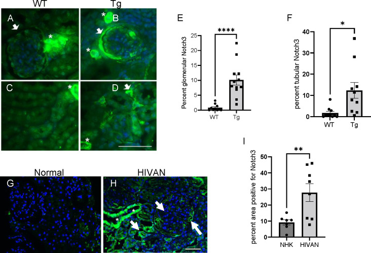
Antiretroviral therapy (ART) has decreased HIV-1 associated morbidity. However, despite ART, immune cells remain latently infected and slowly release viral proteins, leading to chronic inflammation and HIV-1 associated comorbidities. New strategies are needed to target viral proteins and inflammation. We found activation of Notch3 in several renal cells of the HIV-1 mouse model (HIV-Tg26) and in patients with HIV associated Nephropathy. We hypothesized that targeting Notch3 activation constitutes an effective therapy for HIV-related chronic kidney diseases (HIV-CKD). We generated HIV-Tg26 mice with Notch3 knocked out (Tg-N3KO). Compared to HIV-Tg26 mice at 3 months, HIV-Tg-N3KO mice showed a marked reduction in renal injury, skin lesions and mortality rate. Bulk RNA sequencing revealed that N3KO not only reduced renal infiltrating cells but significantly reduced the expression of HIV genes. Moreover, Notch3 activated the HIV- promoter and induction of HIV-1 resulted in increased Notch3 activation indicating a feedback mechanism. Further, bone marrow derived macrophages (BMDMs) from HIV-Tg26 mice showed activation of Notch3 indicating systemic effects. Consistent with that, systemic levels of TNF-α, MCP-1 and other inflammatory chemokines and cytokines were reduced in Tg-N3KO mice. Thus, Notch3 inhibition/deletion has a dual therapeutic effect in HIV-CKD and may extend to other HIV-related pathologies.


Conflict of interest statement

Conflict of interest statement: The authors have declared that no conflict of interest exists.

Figures

Fig. 1.
Fig. 1.. Activation of Notch3 in HIV-1 associated Nephropathy.
(A-B) immunolabeling was performed for presence of Notch3 in renal paraffin sections. Notch3 (green) labeling in kidney sections of 3 months old wildtype (A and C) and Tg26 mice (B and D). Arrows indicate glomerular and tubular interstitial cells highly positive for Notch3 expression. Asterisks indicate blood vessels where Notch3 is normally expressed. (E-F) Quantification of Notch3 expression (intensity, green) as assessed using image J. (G and H) Normal versus HIVAN patient kidney biopsy (representative from n=3 in each group), nuclear expression indicating activation, arrows. (I) Quantification of Notch3 expression (intensity, green) as assessed using image J. Unpaired Student’s t-test was used, and data represented as percent area positive for green labeling (*P<0.05,**P<0.01, ***P<0.001). (Scale bar: 50μm).
Fig. 2.
Fig. 2.. Notch deletion improves disease progression and lifespan in Tg26 mice.
(A) Kaplan Meier curve showing 6 months mortality rate in Tg26 (n=26) and Tg-N3KO (n=28) mice. (B) Phenotypic appearance of WT, N3KO, Tg26 and Tg-N3KO mice, note skin papillomata on the forehead of Tg26 mouse whereas Tg-N3KO mouse appears normal. (C,D) skin lesions/mouse were quantified and area per lesion was measured and expressed as surface area per cm2. (E) Urine was collected in metabolic cages overnight from WT, N3KO, Tg26 and Tg-N3KO mice at 3 months of age before euthanasia. Proteinuria was assessed in 2μl urine by SDS-PAGE electrophoresis followed by Coomassie staining of the gels. “BSA (Bovine serum albumin) was used as a positive control. (F) Albumin and creatinine ratio in urine was measured using ELISA (n=4, WT) (n=4, N3KO) (n=10, Tg26) (n=10,Tg-N3KO) and expressed as μg albumin/mg creatinine. Renal function was also assessed in serum using the blood urea nitrogen (BUN) assay kit and results were expressed as BUN mg/dL (n=4, WT) (n=4, N3KO) (n=6, Tg26) (n=7,Tg-N3KO (E) (*P<0.05) (***P<0.001).
Fig. 3.
Fig. 3.. Notch3 deletion ameliorates kidney injury in Tg26 mice.
(A, B, C) Kidney sections from 3 months old WT, N3KO, Tg26 and Tg-N3KO mice were stained with Periodic acid Schiff (PAS) to determine kidney injury. PAS staining showing tubulointerstitial injury (a), glomerular tubular injury (b) and percentage inflammation (c). Note severe kidney injury in Tg26 kidneys (n=9) as compared to Tg-N3KO (n=12) kidneys. (D, E, F) Blind quantitation of percent tubulointerstitial injury, percent glomerular injury and percentage infiltration (*P<0.05), scale bar 100μm.
Fig. 4.
Fig. 4.. Notch3 targets HIV-1 activity
(A) Heat map showing differential expression of genes obtained from mRNA sequencing of kidneys from 3 months old WT, N3KO, Tg26, Tg-N3KO. Note asterisks for nef and env. (B and C) Quantitative PCR (qPCR) validating nef and env (n=5–7) (D) qPCR to compare Tg-N3KO and Tg-N4KO in repressing nef (n=3 per condition). (E) HIV-LTR promoter luciferase construct and N3IC or PCDNA3.1 (empty vector (EV)) were transiently transfected in podocytes followed by promoter reporter luciferase assays. Data was averaged from 8 assays in 3 independent experiments. Vector transfected control in each experiment was set to one and relative fold change of LTR promoter activity was calculated. (F) Podocytes were transfected with HIV-LTR construct (PNL4) or EV. After 24 hours, lysates were subjected to western blots for N3IC and β-actin . Data was normalized to β-actin via image J and presented as fold change (n=4).
Fig. 5.
Fig. 5.. Notch3 targets immune cell infiltration
(A) Volcano plot comparing gene expression in kidneys from Tg vs WT (top) and Tg-N3KO vs WT (bottom) mice. (B) Immunohistochemistry (IHC) for MMP10 and NFkB-p65 in renal sections of WT, N3KO, Tg26 and Tg-N3KO mice. Arrows show glomerular and tubular areas with high expression. (C) quantitative PCR for ccl2 expression. (D) Serial sections from HIVAN patient biopsy immunostained for the presence of CD68 and Notch3. Note the presence of CD68 positive cells in and around glomerulus, the same areas were also labelled bright for the presence of Notch3 (arrows). (Scale bar: 100μm). (E) Paraffin sections from kidneys of WT, N3KO, Tg26, Tg-N3KO mice, labeled for immune cell markers: CD68, FoxP3 and CD8. (F) Quantification of CD68, FoxP3 and CD8 positive cells from Tg26 and Tg-N3KO mice, blindly assessed in Tg26 (n=9) and Tg-N3KO (n=9) kidneys. Each dot represents percent positive cells from the entire kidney where inflammatory invasions were prominent (*P<0.05), (Scale bar: 50μm). (*P<0.05,**P<0.01, ***P<0.001).
Fig. 6.
Fig. 6.. Abnormal Notch3 expression in bone marrow derived macrophages and systemic inflammation is targeted by Notch3 deletion.
(A) Schematic showing macrophage isolation from bone marrow in mice. (B) Labeling of differentiated bone marrow cells expressing macrophage marker F4/F80 in fixed cells. (C) Lysates obtained from differentiated macrophages of WT and Tg26 mice were subjected to Western blot analysis for N3IC, Dll4, and Jagged1. CD68 was used as a loading control. (D-F) Quantification of bone marrow cells obtained from 3 mice in each group (n=3). (G and H) ELISA assays were performed for the presence of TNFα and MCP-1 in serum obtained from 3 months old WT, N3KO, Tg26 and Tg-N3KO (n=6–12) mice, both males and females. Data is expressed as pg/ml (**P<0.01, ***P<0.001, ****P<0.0001).

References

    1. Gonzales CB, Smith S, Galvan A, Mabry J. The differences between providing oral health care to HIV-infected children and HIV-infected adults: a general dentist’s guide. Gen Dent. Sep-Oct 2010;58(5):424–32; quiz 733–4. - PubMed
    1. Rachakonda AK, Kimmel PL. CKD in HIV-infected patients other than HIV-associated nephropathy. Adv Chronic Kidney Dis. Jan 2010;17(1):83–93. doi:10.1053/j.ackd.2009.09.001 - DOI - PubMed
    1. Papeta N, Sterken R, Kiryluk K, Kalyesubula R, Gharavi AG. The molecular pathogenesis of HIV-1 associated nephropathy: recent advances. J Mol Med (Berl). May 2011;89(5):429–36. doi:10.1007/s00109-010-0719-x - DOI - PubMed
    1. Bachmann N, von Siebenthal C, Vongrad V, et al. Determinants of HIV-1 reservoir size and long-term dynamics during suppressive ART. Nat Commun. Jul 19 2019;10(1):3193. doi:10.1038/s41467-019-10884-9 - DOI - PMC - PubMed
    1. Klatt NR, Chomont N, Douek DC, Deeks SGJIr. Immune activation and HIV persistence: implications for curative approaches to HIV infection. 2013;254(1):326–342. - PMC - PubMed

Publication types