[Preprint]. 2023 Sep 12:2023.09.11.557206.
doi: 10.1101/2023.09.11.557206.

Immune Evasion, Infectivity, and Fusogenicity of SARS-CoV-2 Omicron BA.2.86 and FLip Variants

Affiliations

Immune Evasion, Infectivity, and Fusogenicity of SARS-CoV-2 Omicron BA.2.86 and FLip Variants

Panke Qu et al. bioRxiv. .

Update in

Abstract

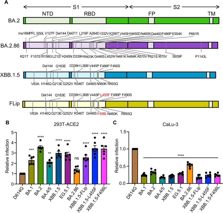
Evolution of SARS-CoV-2 requires the reassessment of current vaccine measures. Here, we characterized BA.2.86 and the XBB-lineage variant FLip by investigating their neutralization alongside D614G, BA.1, BA.2, BA.4/5, XBB.1.5, and EG.5.1 by sera from 3-dose vaccinated and bivalent vaccinated healthcare workers, XBB.1.5-wave infected first responders, and monoclonal antibody (mAb) S309. We assessed the biology of the variant Spikes by measuring viral infectivity and membrane fusogenicity. BA.2.86 is less immune evasive compared to FLip and other XBB variants, consistent with antigenic distances. Importantly, distinct from XBB variants, mAb S309 was unable to neutralize BA.2.86, likely due to a D339H mutation based on modeling. BA.2.86 had relatively high fusogenicity and infectivity in CaLu-3 cells but low fusion and infectivity in 293T-ACE2 cells compared to some XBB variants, suggesting a potentially differences conformational stability of BA.2.86 Spike. Overall, our study underscores the importance of SARS-CoV-2 variant surveillance and the need for updated COVID-19 vaccines.


Conflict of interest statement

Declaration of interests The authors do not declare any competing interests.

Figures

Figure 1:
Figure 1:. Infectivity of Omicron subvariants BA.2.86 and FLip.
(A) Diagrams of the SARS-CoV-2 Omicron subvariants BA.2, BA.2.86, and XBB.1.5 Spikes. The location of specific mutations for BA.2.86 or XBB.1.5 relative to BA.2 in the N-Terminal Domain (NTD) or Receptor Binding Domain (RBD) of the S1 subunit, or in the domain between Fusion Peptide (FP) and Trans-membrane domain (TM) of the S2 subunit, or near the S1/S2 cleavage site is shown. (B-C) Infectivity of pseudotyped lentiviruses bearing each of the indicated Omicron subvariants Spike was determined in (B) HEK293T cells stably expressing human ACE2 (HEK293T-ACE2) or (C) human lung cell-derived epithelial CaLu-3 cells. Transfection efficiency and Spike protein expression were comparable among all groups, which is shown in Figure 5C. Bars in (B-C) represent means ± standard error from triplicates. Significance relative to D614G was analyzed by a one-way repeated measures ANOVA with Bonferroni’s multiple testing correction (n=6). P values are displayed as ns p > 0.05, **p < 0.01, ***p < 0.001, and ****p < 0.0001.
Figure 2:
Figure 2:. Neutralization of Omicron BA.2.86 and FLip subvariants by sera of monovalent or bivalent mRNA vaccinated health care workers (HCWs) and XBB.1.5-wave infection.
Neutralizing antibody (NAb) titers were determined using lentiviruses containing the indicated Spike proteins with D614G as a control. All the NAb titers were compared against D614G. The three cohorts included sera from 14 HCWs who received 3 monovalent doses of mRNA vaccine and 1 dose of bivalent mRNA vaccine (n=14) (A and B), sera from 15 HCWs that received three doses of monovalent mRNA vaccine (n=15) (C and D), and sera from 11 SARS-CoV-2 infected first responders/household contacts or hospitalized patients who tested COVID-19 positive during the XBB1.5- wave of infection in Columbus, Ohio (E and F). Geometric mean NT50 values for each variant are shown on the top. Bars represent geometric means with 95% confidence intervals. Statistical significance was analyzed with log10 transformed NT50 values. Comparisons between multiple groups were performed using a one-way ANOVA with Bonferroni post-test. Dashed lines represent the threshold of detection, i.e., NT50=40. P values are shown as ns p > 0.05, *p < 0.05, **p < 0.01, ***p < 0.001, ****p < 0.0001. Heatmaps in (B, D and F) indicate NAb titers of each individual against each variant tested. Asterisk in (B) indicates that the person being COVID-19 positive within six months before the sera collection, asterisk in (F) indicates that the individuals who received 2 or 3-dose monovalent vaccines before infection, and number sign in (F) indicates that the individuals that received monovalent vaccines and bivalent vaccines.
Figure 3:
Figure 3:. Neutralization of Omicron BA.2.86 and FLip subvariants by monoclonal antibody (mAb) S309.
The lentiviral pseudotypes carrying each of the indicated variant Spike proteins were used to assess the effectiveness of mAb S309 in neutralizing BA.2.86, Flip and other variants. Representative plot curves are displayed (A) and the calculated IC50 values (means ± standard deviation) from two biological replicates are shown (B).
Figure 4:
Figure 4:. Cell-cell fusion of Omicron BA.2.86 and FLip subvariants.
HEK293T cells were cotransfected with the indicated Spikes of interest and GFP plasmids, and were cocultured with HEK29T cells overexpressing human ACE2 (293T-ACE2) (A-B) or human lung epithelial CaLu-3 cells (C-D) for 24 hours. Cell-cell fusion was imaged and GFP areas of fused cells were quantified (see Methods). D614G and no S were included as positive and negative control, respectively. Comparisons in extents of cell-cell fusion for each Omicron subvariant were made against D614G. Bars in (B and D) represent means ± standard error. Dots represent three images from two biological replicates. Statistical significance relative to D614G was determined using a one-way repeated measures ANOVA with Bonferroni’s multiple testing correction (n=3). P values are displayed as ns p > 0.05, *p <0.05, ***p < 0.001, and ****p < 0.0001.
Figure 5:
Figure 5:. Cell surface expression and processing of Omicron BA.2.86 and Flip Spike proteins.
(A-B) Cell surface expression of the indicated variant Spike proteins. HEK293T cells used for production of pseudotyped lentiviral vectors carrying each variant Spike proteins (Figures 1–3) were stained with anti-SARS-CoV-2 S1 antibody. Representative histogram of anti-S1 signals in the cells (A) and geometric mean fluorescence intensities (B) of each subvariant from three biological replicates are shown. (C) Spike expression and processing in viral producer cell lysates. HEK293T cells, which were used to produce lentiviral pseudotypes, were lysed and probed with anti-S1, anti-S2 and anti-GAPDH antibodies, respectively. Spike processing was quantified by NIH ImageJ and set to a S1/S or S2/S ratio and normalized the ratios of each Omicron subvariant to that of D614G. Dots represent three biological replicates. Bars in (B) represent means ± standard error. Significance relative to D614G was made using a one-way ANOVA with Bonferroni post-test. P values are displayed as ns p > 0.05, *p < 0.05, **p < 0.01, and ***p < 0.001.
Figure 6:
Figure 6:. Homology modeling of key mutations in BA.2.86.
(A) A homology model of the BA.2.86 Spike trimer is presented, highlighting mutations that differ from the BA.2 variant as red sticks on the green protomer. (B) The substitution of the wildtype G339 residue with either D or H introduces steric hindrance to residues Y100 and L110 of antibody S309. Simultaneously, the K356T mutation disrupts the salt-bridge interaction with E108 of S309. These mutations collectively impair the recognition of the Spike protein by antibody S309. (C) The A570V mutation in BA.2.86 Spike enhances hydrophobic interactions between protomers, thereby increasing trimer stability. (D) Focusing on the RBM region, the V445H and R493Q mutations may enhance receptor binding by introducing hydrogen bonds between the Spike protein and the ACE2 receptor. Conversely, the F486P mutation weakens receptor binding by losing the hydrophobic interaction with F83 of ACE2.
Figure 7:
Figure 7:. Antigenic mapping of neutralization titers for bivalent vaccinated, monovalent vaccinated, and XBB.1.5-wave infected cohorts, derived from the NT50 in Figure 2.
Antigenic maps for neutralization titers from (A) the bivalent vaccinated, (B) the monovalent vaccinated, and (C) the XBB.1.5-wave infected cohorts were made using the Racmacs program (1.1.35) (see Methods). Squares represent the individual sera sample and circles represent the variants. One square on the grid represents one antigenic unit squared.

References

    1. Gangavarapu K., Latif A., Mullen J., Alkuzweny M., Hufbauer E., Tsueng G., Haag E., Zeller M., Aceves C., and Zaiets K. (2023). SARS-CoV-2 (hCoV-19) Mutation Reports. - PMC - PubMed
    1. Yuan S., Ye Z.-W., Liang R., Tang K., Zhang A.J., Lu G., Ong C.P., Man Poon V.K., Chan C.C.-S., Mok B.W.-Y., et al. (2022). Pathogenicity, transmissibility, and fitness of SARS-CoV-2 Omicron in Syrian hamsters. Science 377, 428–433. doi: 10.1126/science.abn8939. - DOI - PubMed
    1. Suzuki R., Yamasoba D., Kimura I., Wang L., Kishimoto M., Ito J., Morioka Y., Nao N., Nasser H., Uriu K., et al. (2022). Attenuated fusogenicity and pathogenicity of SARS-CoV-2 Omicron variant. Nature 603, 700–705. 10.1038/s41586-022-04462-1. - DOI - PMC - PubMed
    1. Shuai H., Chan J.F., Hu B., Chai Y., Yuen T.T., Yin F., Huang X., Yoon C., Hu J.C., Liu H., et al. (2022). Attenuated replication and pathogenicity of SARS-CoV-2 B.1.1.529 Omicron. Nature 603, 693–699. 10.1038/s41586-022-04442-5. - DOI - PubMed
    1. Zeng C., Evans J.P., Qu P., Faraone J., Zheng Y.M., Carlin C., Bednash J.S., Zhou T., Lozanski G., Mallampalli R., et al. (2021). Neutralization and Stability of SARS-CoV-2 Omicron Variant. bioRxiv. 10.1101/2021.12.16.472934. - DOI

Publication types