[Preprint]. 2023 Sep 15:2023.09.12.557419.
doi: 10.1101/2023.09.12.557419.

The αC-β4 loop controls the allosteric cooperativity between nucleotide and substrate in the catalytic subunit of protein kinase A

Affiliations

The αC-β4 loop controls the allosteric cooperativity between nucleotide and substrate in the catalytic subunit of protein kinase A

Cristina Olivieri et al. bioRxiv. .

Update in

Abstract

Allosteric cooperativity between ATP and substrates is a prominent characteristic of the cAMP-dependent catalytic (C) subunit of protein kinase A (PKA). Not only this long-range synergistic action is involved in substrate recognition and fidelity, but it is likely to regulate PKA association with regulatory subunits and other binding partners. To date, a complete understanding of the molecular determinants for this intramolecular mechanism is still lacking. Here, we used an integrated NMR-restrained molecular dynamics simulations and a Markov Model to characterize the free energy landscape and conformational transitions of the catalytic subunit of protein kinase A (PKA-C). We found that the apo-enzyme populates a broad free energy basin featuring a conformational ensemble of the active state of PKA-C (ground state) and other basins with lower populations (excited states). The first excited state corresponds to a previously characterized inactive state of PKA-C with the αC helix swinging outward. The second excited state displays a disrupted hydrophobic packing around the regulatory (R) spine, with a flipped configuration of the F100 and F102 residues at the tip of the αC-β4 loop. To experimentally validate the second excited state, we mutated F100 into alanine and used NMR spectroscopy to characterize the binding thermodynamics and structural response of ATP and a prototypical peptide substrate. While the activity of PKA-CF100A toward a prototypical peptide substrate is unaltered and the enzyme retains its affinity for ATP and substrate, this mutation rearranges the αC-β4 loop conformation interrupting the allosteric coupling between nucleotide and substrate. The highly conserved αC-β4 loop emerges as a pivotal element able to modulate the synergistic binding between nucleotide and substrate and may affect PKA signalosome. These results may explain how insertion mutations within this motif affect drug sensitivity in other homologous kinases.

Keywords: Protein Kinases; allosteric mutations; binding cooperativity; cAMP-dependent protein kinase A.


Conflict of interest statement

Competing interest The authors declare no financial and non-financial competing interests.

Figures

Figure 1.
Figure 1.. Structural and catalytic motifs of PKA-C.
(A) Surface representation of the X-ray structure of PKA-C bound to the endogenous inhibitor, PKI (PDB: 4WB5). (B) Hydrophobic organization of the PKA-C core, with the R-spine (gold), C-spine (blue), shell residues (cyan), and the αC-β4 loop (hot pink) that locks into αE helix.
Figure 2.
Figure 2.. Free energy landscape (FEL) of PKA-C in various ligated forms obtained from replica-averaged metadynamics (RAM) simulations.
(A) Convergence of the bias deposition along the first three collective variables (CVs). The free energy (expressed in kcal/mol) of the different CVs were averaged over the last 100 ns of RAM simulations. The standard deviations are reported as red error bars. (B-D) FEL along the first two principal components (PC1 and PC2) of PKA-C in the apo, ATP-bound, and ATP and the model substrate PKI bound forms. PC1 and PC2 are projected from the first three CVs. The vertices represent conformational states. In the apo form, multiple states have comparable free energy with ∆G < 5 kcal/mol, whereas in the binary form, fewer states have ∆G < 5 kcal/mol, whereas for the ternary form only a major ground state is populated.
Figure 3.
Figure 3.. The apo, ATP-bound, and ATP/PKI-bound PKA-C reveal distinct free energy surface (FES) and dynamics, as determined by a Markov State Model (MSM).
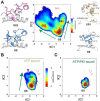
(A) Free energy landscape projected along the first two time-lagged independent components (tICs) of the apo PKA-C, the projections of known crystal structures, and characteristic features of GS, ES1, and ES2. The transition from GS to ES1 highlights the changes around the αB-αC loop, where the salt bridges between K72-E91 and H87-T197 and the PIF pocket (V80-I85-F347) are all disrupted. The transition from GS to ES2 highlights the rearrangement around the αC-β4 loop, with distinct local hydrophobic packing. (B and C) FES projected along the first two tICs for the ATP-bound PKA-C (B), ATP/PKI bound PKA-C (C), and the projections of known crystal structures.
Figure 4.
Figure 4.. Conformational transition between GS and ES1, ES2 along the kinetic Monte Carlo trajectory in apo (A) and ATP-bound (B) forms.
(A, B) The transition from GS to ES1, revealed as breaking of the K72-E91 salt bridge, is frequently found in both forms, whereas the transition to ES2, revealed as the contact between F100 and V104, only occurs in the apo form and in concert with allosteric changes between D166-N171, K168-T201, and W222-A206-P207. The darker colors in (A) and (B) highlight moving averages over every 10 frames. (C) GS conformation reveals the assembly of key catalytic features across the core region. (D) ES2 conformation revealed disruption of key structure motifs across the core region, indicative of inactivation.
Figure 5.
Figure 5.. Transition from GS to ES2 shown in the apo PKA-C recapitulated the structural changes near the αC- β4 loop probed by NMR experiments.
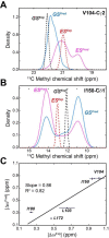
(A and B) Distribution of predicted 13C CS of selected methyl groups in ES (magenta) and GS (blue) of the apo PKA-C for Val104-Cγ1 (A) and Ile150-Cδ1 (B). The experimental CS is shown in dotted lines for GS (black) and ES (red). (C). Correlation of the predicted chemical shift differences | ∆ωPred| and the experimental result | ∆ωExp| for a set of hydrophobic residues near the αC-β4 loop. The fitted linear correlation has a slope of 0.86 and R2 of 0.82.
Figure 6.
Figure 6.. Increased dynamics at the αC-β4 loop upon F100A mutation, perturbing the local hydrophobic packing and its anchoring to the αE helix.
(A) Time series of the αC-β4 loop, H-bond occurrence for the β– and γ-turns, F102 χ1 angle, and N99 and Y156 for WT (black) and F100A (red) in the ATP-bound state. (B) Representative structural snapshots showing the formation of the β-turn for the PKA-CWT (green) and γ-turn for the PKA-CF100A mutant.
Figure 7.
Figure 7.. Distinct global structural response to ATP binding in the F100A mutant.
(A) Structure superposition of the C Spine, R Spine, and Shell residues between WT (lime) and F100A (hot pink), highlighting the differences between Shell and R Spine. (B) Change of RMSD upon ATP binding at C Spine, Shell, and R spine, for WT and F100A, respectively. (C) Structural illustration of the first principal component (PC1), i.e., the breathing motion of the two lobes. (D) Structural illustration of the second principal component (PC2), i.e., the shearing motion of the two lobes. (E) Comparison of the 2D projection and distribution along PC1 and PC2 for WT and F100A, highlighting their dramatic differences along both axes.
Figure 8.
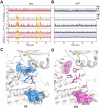
Figure 8.. Mutual information of dihedral angles for the (A) WT and (B) F100A upon binding ATP.
This analysis reveals the prominent loss of allosteric communication of F100A, especially at multiple key motifs as is highlighted by purple strips.
Figure 9.
Figure 9.. Structural response of PKA-CF100A binding to nucleotide and protein kinase inhibitor.
(A) Histogram shows the chemical shift perturbation (CSP) of the amide fingerprint for PKA-CF100A (black) in response to ATPγN binding compared to the CSP obtained for the wild-type protein (cyan). The dashed line on the histogram indicates one standard deviation from the average CSP. (B) CSPs of PKA-CF100A/ATPγN amide resonances mapped onto the structure (PDB: 4WB5). (C) CSP of amide fingerprint for PKA-CF100A bound to ATPγN and PKI5–24 (black), compared to the CSP of the wild-type protein obtained in the same conditions. (D) CSP for the F100A/ATPγN/PKI complex mapped onto the crystal structure (PDB: 4WB5).
Figure 10 –
Figure 10 –. Changes of the intramolecular allosteric network in F100A as mapped by correlated chemical shift changes.
(A) Comparison of the CHESCA matrices obtained from the analysis of the amide chemical shifts of PKA-CWT (top diagonal, blue) and PKA-CF100A (bottom diagonal, black) in the apo, ADP-bound, ATPγN-bound, and ATPγN/PKI5–24-bound states. Only correlations with Rij > 0.98 are reported. The enlarged CHESCA map of F100A is available in Figure 10 – figure supplement 1 while the data for the PKA-CWT matrix are taken from Walker et al.. (B) Community CHESCA analysis of PKA-CWT (top diagonal, blue) and PKA-CF100A (bottom diagonal, black). Only correlations with RA,B > 0.98 are shown. (C) Community CHESCA matrix plotted on its corresponding structures. The size of each node is independent of the number of residues it encompasses while the weight of each line indicates the strength of coupling between the individual communities.

References

    1. Manning G., Whyte D.B., Martinez R., Hunter T. & Sudarsanam S. The Protein Kinase Complement of the Human Genome. Science 298, 1912–1934 (2002). - PubMed
    1. Knighton D.R. et al. Crystal structure of the catalytic subunit of cyclic adenosine monophosphate-dependent protein kinase. Science 253, 407–14 (1991). - PubMed
    1. Knighton D.R. et al. Structure of a peptide inhibitor bound to the catalytic subunit of cyclic adenosine monophosphate-dependent protein kinase. Science 253, 414–20 (1991). - PubMed
    1. Taylor S.S., Ilouz R., Zhang P. & Kornev A.P. Assembly of allosteric macromolecular switches: lessons from PKA. Nat Rev Mol Cell Biol 13, 646–58 (2012). - PMC - PubMed
    1. Walsh D.A., Perkins J.P. & Krebs E.G. An adenosine 3',5'-monophosphate-dependant protein kinase from rabbit skeletal muscle. J Biol Chem 243, 3763–5 (1968). - PubMed

Publication types