Case Reports
. 2023 Sep 14;18(11):4130-4133.
doi: 10.1016/j.radcr.2023.08.080. eCollection 2023 Nov.

Odontogenic myxoma: A case report of a rare tumor

Affiliations
Case Reports

Odontogenic myxoma: A case report of a rare tumor

Andrés Felipe Herrera Ortiz et al. Radiol Case Rep. .

Abstract

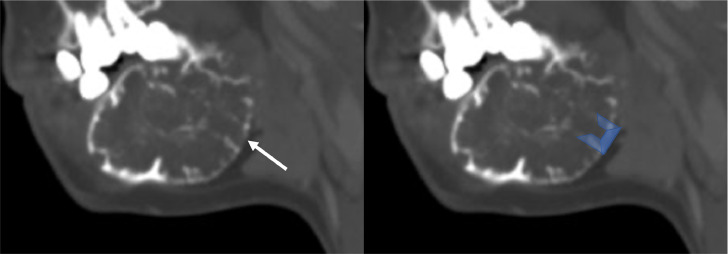
Odontogenic myxoma is a rare and aggressive tumor. Identifying the tumor based on imaging characteristics can pose a challenge due to similarities in features with other tumors, such as ameloblastomas and aneurysmal bone cysts. We report a 33-year-old female who presented with a palpable, tender mass in the lower right jaw. A computed tomography scan revealed a multicystic tumor which was proved to be an odontogenic myxoma. The patient underwent partial surgical resection followed by CO2 laser-assisted evaporation. During 1-year follow-up, the patient showed satisfactory results and no signs of tumor growth. This case report highlights the diagnostic challenges associated with odontogenic myxoma, emphasizing age as a key diagnostic feature.

Keywords: Ameloblastoma; Aneurysmal bone cyst; Computed tomography; Jaw tumor; Odontogenic myxoma.


Figures

Fig 1
Fig. 1
Noncontrast CT in bone window (A–C) and volume rendering reconstruction (D). The images show a multiloculated lytic lesion located in the body and angle of the right mandible, causing expansion and thinning of the adjacent cortical bone (white arrow); note that the tumor is in intimate contact with the right inferior second premolar teeth (orange). Collectively, these findings are consistent with odontogenic myxoma; however other differential diagnoses, such as ameloblastoma and aneurysmal bone cyst, are feasible.
Fig 2
Fig. 2
Bone window CT with sagittal reconstruction. The image shows fine septa perpendicular to the lesion's borders, separating the tumor into rectangular spaces (white arrow and blue U shape).
Fig 3
Fig. 3
Noncontrast CT in bone window (A–C) and volume rendering reconstruction (D). The images show an area of curettage in the body of the mandible due to marginal mandibulectomy (white arrows), surrounded by multiloculated lytic lesions consistent with residual tumor that could not be removed (orange arrow).

References

    1. Reddy SP, Naag A, Kashyap B. Odontogenic myxoma: report of two cases. Natl J Maxillofac Surg. 2010;1(2):183–186. doi: 10.4103/0975-5950.79227. - DOI - PMC - PubMed
    1. Simon ENM, Merkx MAW, Vuhahula E, Ngassapa D, Stoelinga PJW. Odontogenic myxoma: a clinicopathological study of 33 cases. Int J Oral Maxillofac Surg. 2004;33(4):333–337. doi: 10.1016/j.ijom.2003.12.004. - DOI - PubMed
    1. Shivashankara C, Nidoni M, Patil S, Shashikala KT. Odontogenic myxoma: a review with report of an uncommon case with recurrence in the mandible of a teenage male. Saudi Dent J. 2017;29(3):93–101. doi: 10.1016/j.sdentj.2017.02.003. - DOI - PMC - PubMed
    1. Wang K, Guo W, You M, Liu L, Tang B, Zheng G. Characteristic features of the odontogenic myxoma on cone beam computed tomography. Dentomaxillofac Radiol. 2017;46(2) doi: 10.1259/dmfr.20160232. - DOI - PMC - PubMed
    1. Fenton S, Slootweg PJ, Dunnebier EA, Mourits MP. Odontogenic myxoma in a 17-month-old child: a case report. J Oral Maxillofac Surg. 2003;61(6):734–736. doi: 10.1053/joms.2003.50121. - DOI - PubMed

Publication types

LinkOut - more resources