. 2023 Sep 18:16:4103-4116.
doi: 10.2147/JIR.S416239. eCollection 2023.

HDAC1 is Involved in Neuroinflammation and Blood-Brain Barrier Damage in Stroke Pathogenesis

Affiliations

HDAC1 is Involved in Neuroinflammation and Blood-Brain Barrier Damage in Stroke Pathogenesis

Hao-Kuang Wang et al. J Inflamm Res. .

Abstract

Background: Stroke is a common cause of disability and mortality worldwide; however, effective therapy remains limited. In stroke pathogenesis, ischemia/reperfusion injury triggers gliosis and neuroinflammation that further activates matrix metalloproteinases (MMPs), thereby damaging the blood-brain barrier (BBB). Increased BBB permeability promotes macrophage infiltration and brain edema, thereby worsening behavioral outcomes and prognosis. Histone deacetylase 1 (HDAC1) is a repressor of epigenomic gene transcription and participates in DNA damage and cell cycle regulation. Although HDAC1 is deregulated after stroke and is involved in neuronal loss and DNA repair, its role in neuroinflammation and BBB damage remains unknown.

Methods: The rats with cerebral ischemia were evaluated in behavioral outcomes, levels of inflammation in gliosis and cytokines, and BBB damage by using an endothelin-1-induced rat model with cerebral ischemia/reperfusion injury.

Results: The results revealed that HDAC1 dysfunction could promote BBB damage through the destruction of tight junction proteins, such as ZO-1 and occludin, after stroke in rats. HDAC1 inhibition also increased the levels of astrocyte and microglial gliosis, tumor necrosis factor-alpha, interleukin-1 beta, lactate dehydrogenase, and reactive oxygen species, further triggering MMP-2 and MMP-9 activity. Moreover, modified neurological severity scores for the cylinder test revealed that HDAC1 inhibition deteriorated behavioral outcomes in rats with cerebral ischemia.

Discussion: HDAC1 plays a crucial role in ischemia/reperfusion-induced neuroinflammation and BBB damage, thus indicating its potential as a therapeutic target.

Keywords: HDAC1; IL-1β; ROS; TNF-α; blood-brain barrier; cylinder test; mNSS; stroke.


Conflict of interest statement

The authors declare there is no conflict of interest in this study.

Figures

Figure 1
Figure 1
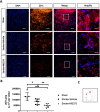
HDAC1 dysfunction promoted BBB damage in rats 24 h after stroke. (A) The representative data of Evan`s blue staining from the cerebral ischemia rats 24 h after surgery. (B) The quantified data for Evan`s blue staining from cerebral ischemia rats. Sham n=6, Stroke+Vehicle n=8, Stroke+MS275 n=8. Data was evaluated by one-way ANOVA, *p < 0.05, **p <0.01, ***p <0.001.
Figure 2
Figure 2
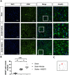
HDAC1 dysfunction decreased the expression of tight junction associated protein- ZO-1 24 h after stroke. (A) The representative figures of immunofluorescent staining for ZO-1 in cerebral ischemia rats 24 h after stroke. The white square denotes an amplified view from the merged figure. Bar: 100 μm; Bar: 400 μm in amplify (B) The quantified data of immunofluorescent staining. (C) The red square denotes where the general view of immunostainings was captured in the brain sections. Sham n=6, Stroke+Vehicle n=8, Stroke+MS275 n=8. Data was evaluated by one-way ANOVA, *p < 0.05, **p <0.01, ***p <0.001.
Figure 3
Figure 3
HDAC1 dysfunction reduced the tight junction-associated proteins in ZO-1 and occluding 24 h after stroke. (A) The representative Western blotting data for ZO-1 and occludin 24 h after stroke. β-actin served as an internal control. (B) The quantified levels of ZO-1 and occluding normalized to internal control. N=6 per group. Data was evaluated by one-way ANOVA, *p < 0.05, **p < 0.01, ***p < 0.001.
Figure 4
Figure 4
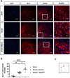
HDAC1 dysfunction increased the astrocytic gliosis 24 h after stroke. (A) The representative figures of immunofluorescent staining for GFAP in cerebral ischemia rats. The white square denotes an amplified view from the merged figure. Bar: 50 μm; Bar: 200 μm in amplify (B) The quantified data of immunofluorescent staining for GFAP. (C) The red square denotes where the general view of immunostainings was captured in the brain sections. Sham n=6, Stroke+Vehicle n=8, Stroke+MS275 n=8. Data was evaluated by one-way ANOVA, *p < 0.05, ***p <0.001.
Figure 5
Figure 5
HDAC1 dysfunction increased the microglial gliosis 24 h after stroke. (A) The representative figures of immunofluorescent staining for GFAP in cerebral ischemia rats. The white square denotes an amplified view from the merged figure. Bar: 50 μm; Bar: 200 μm in amplify (B) The quantified data of immunofluorescent staining for GFAP. (C) The red square denotes where the general view of immunostainings was captured in the brain sections. Sham n=6, Stroke+Vehicle n=8, Stroke+MS275 n=8. Data was evaluated by one-way ANOVA, *p < 0.05, **p <0.01, ****p <0.0001.
Figure 6
Figure 6
HDAC1 dysfunction worsened the levels of inflammation cytokines, ROS, and LDH 24h after stroke. (A and B) ELISA was conducted for inflammation cytokines in IL-1b and TNF-a. (C and D) ROS and LDH were detected 24 h after stroke from the brain lysates of cerebral ischemia rats. (E) MMPs activity assay was conducted to evaluate the enzymatic activity of MMP2 and MMP9 24 h after stroke. Sham n=6, Stroke+Vehicle n=8, Stroke+MS275 n=8. Data was evaluated by one-way ANOVA, *p < 0.05, **p <0.01, ***p <0.001.
Figure 7
Figure 7
HDAC1 dysfunction elicited worsened behavioral outcomes in the mNSS and cylinder tests at post-stroke days (PSD) 1, 3, 7. (A) Evaluations for neuromuscular function by modified neural severity scores (mNSS) in cerebral ischemia rats at PSD 1, 3, 7. (B) Evaluation for forepaw using ability in cerebral ischemia rats at PSD 1, 3, 7. Sham n=6, Stroke+Vehicle n=8, Stroke+MS275 n=8. Data was evaluated by one-way ANOVA, *p < 0.05, **p <0.01.

Similar articles

Cited by

References

    1. Kuriakose D, Xiao Z. Pathophysiology and treatment of stroke: present status and future perspectives. Int J Mol Sci. 2020;21(20):7609. doi:10.3390/ijms21207609 - DOI - PMC - PubMed
    1. Feigin VL, Stark BA, Johnson CO, et al. Global, regional, and national burden of stroke and its risk factors, 1990-2019: a systematic analysis for the global burden of disease study 2019. Lancet Neurol. 2021;20(10):795–820. doi:10.1016/S1474-4422(21)00252-0 - DOI - PMC - PubMed
    1. Donkor ES. Stroke in the 21(st) century: a snapshot of the burden, epidemiology, and quality of life. Stroke Res Treat. 2018;2018:3238165. doi:10.1155/2018/3238165 - DOI - PMC - PubMed
    1. Katan M, Luft A. Global burden of stroke. Semin Neurol. 2018;38(2):208–211. doi:10.1055/s-0038-1649503 - DOI - PubMed
    1. Profaci CP, Munji RN, Pulido RS, Daneman R. The blood-brain barrier in health and disease: important unanswered questions. J Exp Med. 2020;217(4). doi:10.1084/jem.20190062 - DOI - PMC - PubMed