. 2020 Aug;2(3):e202000004.
doi: 10.1002/tbio.202000004. Epub 2020 May 12.

Highly sensitive lipid detection and localization in atherosclerotic plaque with a dual-frequency intravascular photoacoustic/ultrasound catheter

Affiliations

Highly sensitive lipid detection and localization in atherosclerotic plaque with a dual-frequency intravascular photoacoustic/ultrasound catheter

Yingchun Cao et al. Transl Biophotonics. 2020 Aug.

Abstract

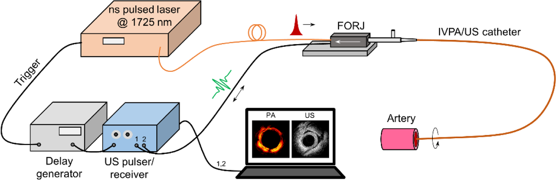
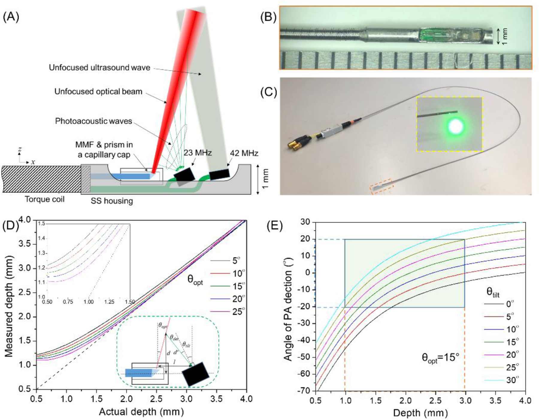
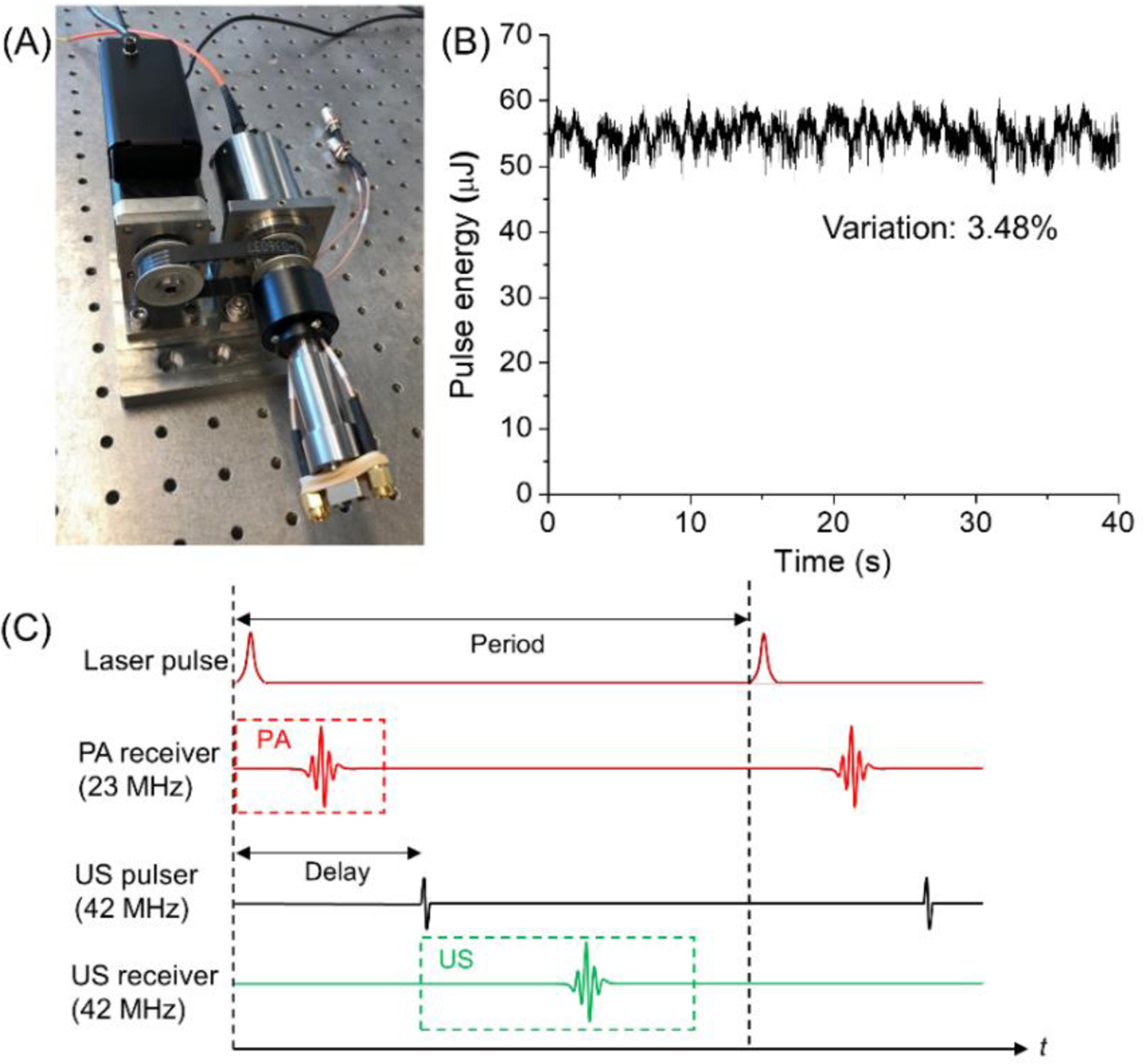
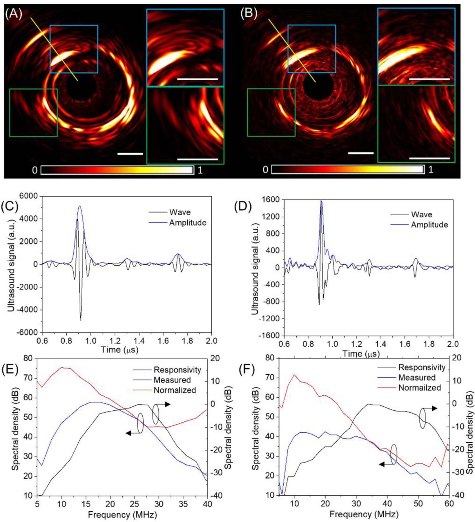
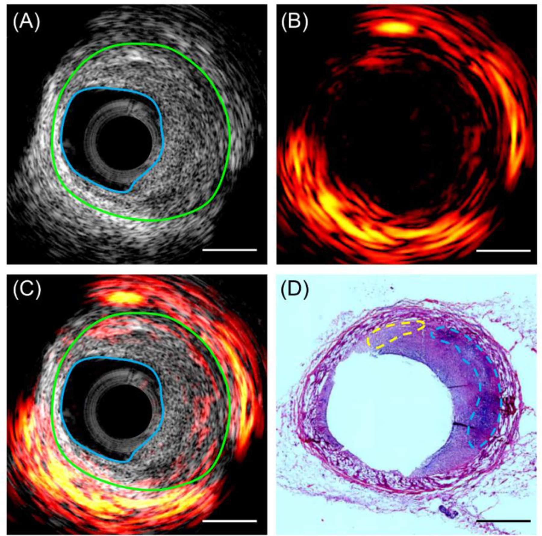
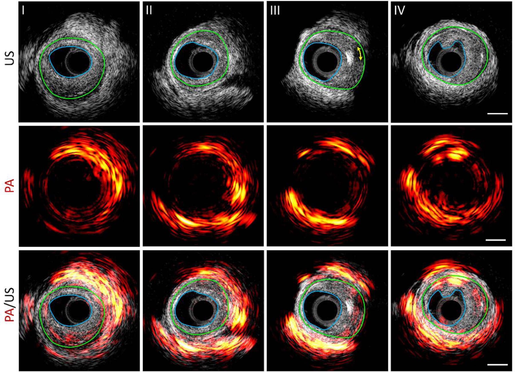
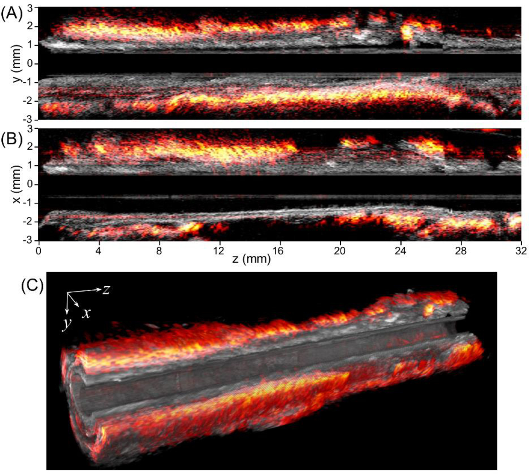
Intravascular photoacoustic/ultrasound (IVPA/US) is an emerging hybrid imaging modality that provides specific lipid detection and localization, while maintaining co-registered artery morphology, for diagnosis of vulnerable plaque in cardiovascular disease. However, current IVPA/US approaches based on a single-element transducer exhibit compromised performance for lipid detection due to the relatively low contrast of lipid absorption and conflicting detection bands for photoacoustic and ultrasound signals. Here, we present a dual-frequency IVPA/US catheter for highly sensitive detection and precision localization of lipids. The low frequency transducer provides enhanced photoacoustic sensitivity, while the high frequency transducer maintains state-of-the-art spatial resolution for ultrasound imaging. The boosted capability of IVPA/US imaging enables a multi-scale analysis of lipid distribution in swine with coronary atherosclerosis. The dual-frequency IVPA/US catheter has a diameter of 1 mm and flexibility to easily adapt to current catheterization procedures and is a significant step toward clinical diagnosis of vulnerable plaque.

Keywords: Ossabaw miniature swine; atherosclerotic plaque; dual-frequency; intravascular photoacoustic; lipid core.


Conflict of interest statement

CONFLICT OF INTEREST The authors declare no potential conflict of interests.

Figures

FIGURE 1.
FIGURE 1.
[Schematic of dual-frequency IVPA/US imaging. FORJ: fiber-optic rotary joint.]
FIGURE 2.
FIGURE 2.
[Dual-frequency IVPA/US catheter design and optimization. (A) Design of a dual-frequency IVPA/US catheter; (B) photograph of fabricated catheter tip; (C) entire catheter; (D) optimization of optical firing angle from the catheter tip, with the upper inset showing a zoom-in at small depth, and the lower inset depicting the geometrical relationship between fiber tip and 23 MHz transducer; (E) optimization of tilted angle of 23 MHz transducer with light firing angle of 15° to the vertical direction, the green area showing the effective detection region and acceptance angle of the transducer.]
FIGURE 3.
FIGURE 3.
[Optical and electrical coupling, and time sequence design. (A) Photograph of a hybrid fiber-optic & electrical rotary joint; (B) coupled optical pulse energy monitoring during continuous 10 revolutions; (C) strategy of time sequence design of the two transducer channels.]
FIGURE 4.
FIGURE 4.
[Comparison between 23 MHz and 42 MHz transducers for detection of lipid deposit in artery using a collinear catheter design. (A, B) Normalized photoacoustic images with zoom-in display of selected regions; (C, D) A-line signal and amplitude along the yellow line in (A, B); (E, F) frequency response of photoacoustic signals in (C, D). The black lines in (E, F) are responsivity of transducers characterized by measuring the ultrasound echo from a rigid surface, the blue lines are frequency domain expression of measured signal, while the red lines are their normalized result to the responsivity. (A, C, E) correspond to 23 MHz catheter, and (B, D, F) are for 42 MHz transducer.]
FIGURE 5.
FIGURE 5.
[Representative imaging results and histology validation. (A) Ultrasound, (B) photoacoustic, (C) their overlaid resulting image at a representative cross-sectional location of the pig artery. The region between the blue line and green line indicates a thickened intima, i.e. atherosclerotic plaque; (D) hematoxylin and eosin histology result at the same artery location. The blue line indicates lipid core, while the yellow lines denotes its early-stage lipid deposits. Scale bar of 1 mm applies to all the panels.]
FIGURE 6.
FIGURE 6.
[Photoacoustic (PA), ultrasound (US), and their overlaid images at representative positions along a pig coronary artery. Scale bar of 1 mm (right column) applies to all the panels. The blue line denotes the boundary of lumen and the green line indicates the boundary of the neo-intima layer (boundary of initial lumen). The yellow arrowed line in III indicates the acoustic shadowing behind an echogenic calcium nodule.]
FIGURE 7.
FIGURE 7.
[2D and 3D overlaid photoacoustic/ultrasound images over the pullback of the IVPA/US transducer through 32 mm length of artery. (A, B) Overlaid photoacoustic/ultrasound images in yz and xz planes, respectively, (C) 3D photoacoustic/ ultrasound images of the artery with a 24 mm section length and a quarter cutaway for better visualization.]

Similar articles

Cited by

References

    1. Falk E, Shah Prediman K, Fuster V, Circulation 1995, 92(3), 657–671. - PubMed
    1. Virmani R, Burke AP, Kolodgie FD, Farb A, J. Interv. Cardiol 2003, 16(3), 267–272; - PubMed
    2. Virmani R, Burke AP, Farb A, Kolodgie FD, J. Am. Coll. Cardiol 2006, 47(8), C13–C18. - PubMed
    1. Wahle A, Lopez JJ, Olszewski ME, Vigmostad SC, Chandran KB, Rossen JD, Sonka M, Med. Image Anal 2006, 10(4), 615–631. - PMC - PubMed
    1. Tarkin JM, Dweck MR, Evans NR, Takx RAP, Brown AJ, Tawakol A, Fayad ZA, Rudd JHF, Circ. Res 2016, 118(4), 750–769. - PMC - PubMed
    1. Wang Z, Jiang X, Czernuszewicz TJ, Gallippi CM, Dual-frequency IVUS transducer for acoustic radiation force impulse (ARFI) imaging, in 2015 IEEE International Ultrasonics Symposium (IUS), IEEE, 2015, pp. 1–4;
    2. Shih C-C, Chen P-Y, Ma T, Zhou Q, Shung KK, Huang C-C, R. Soc. Open Sci 2018, 5(4), 180138–180138. - PMC - PubMed

LinkOut - more resources