

Incidents of snake fungal disease caused by the fungal pathogen Ophidiomyces ophidiicola in Texas
- PMID: 37746129
- PMCID: PMC10512329
- DOI: 10.3389/ffunb.2023.1064939
Incidents of snake fungal disease caused by the fungal pathogen Ophidiomyces ophidiicola in Texas
Abstract
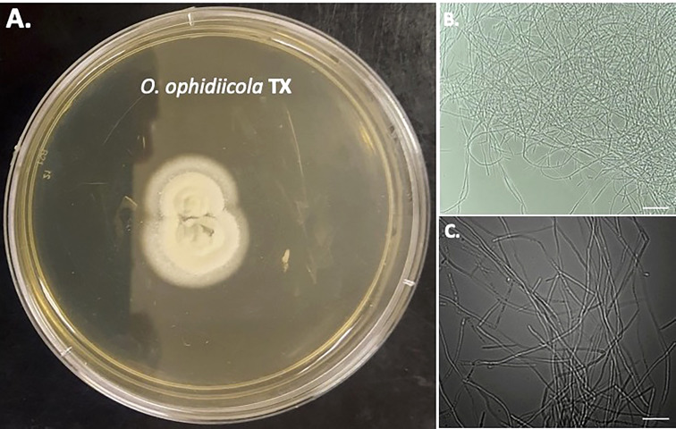
The pathogen Ophidiomyces ophidiicola, widely known as the primary cause of snake fungal disease (SFD) has been detected in Texas's naïve snakes. Our team set out to characterize O. ophidiicola's spread in eastern Texas. From December 2018 until November 2021, we sampled and screened with ultraviolet (UV) light, 176 snakes across eastern Texas and detected 27. O. ophidiicola's positive snakes using qPCR and one snake in which SFD was confirmed via additional histological examination. Upon finding the ribbon snake with clear clinical display, we isolated and cultured what we believe to be the first culture from Texas. This cultured O. ophidiicola TX displays a ring halo formation when grown on a solid medium as well as cellular autofluorescence as expected. Imaging reveals individual cells within the septated hyphae branches contain a distinct nucleus separation from neighboring cells. Overall, we have found over 1/10 snakes that may be infected in East Texas, gives credence to the onset of SFD in Texas. These results add to the progress of the disease across the continental United States.
Keywords: Ophidiomyces ophidiicola; Texas; UV fluorescence; fungal Infection; snake fungal disease.
Copyright © 2023 Lizarraga, Hart, Wright, Williams and Glavy.
Conflict of interest statement
The authors declare that the research was conducted in the absence of any commercial or financial relationships that could be construed as a potential conflict of interest.
Figures


Similar articles
-
Testing the febrile response of snakes inoculated with Ophidiomyces ophidiicola, the causative agent of snake fungal disease.J Therm Biol. 2021 Aug;100:103065. doi: 10.1016/j.jtherbio.2021.103065. Epub 2021 Aug 3. J Therm Biol. 2021. PMID: 34503803
-
Prevalence of Ophidiomyces ophidiicola and epizootiology of snake fungal disease in free-ranging Northern Pine Snakes (Pituophis melanoleucus melanoleucus) in New Jersey.Environ Monit Assess. 2023 May 11;195(6):662. doi: 10.1007/s10661-023-11259-w. Environ Monit Assess. 2023. PMID: 37169998
-
Environmental associations of Ophidiomyces ophidiicola, the causative agent of ophidiomycosis in snakes.PLoS One. 2024 Oct 22;19(10):e0310954. doi: 10.1371/journal.pone.0310954. eCollection 2024. PLoS One. 2024. PMID: 39436883 Free PMC article.
-
Snake fungal disease: an emerging threat to wild snakes.Philos Trans R Soc Lond B Biol Sci. 2016 Dec 5;371(1709):20150457. doi: 10.1098/rstb.2015.0457. Philos Trans R Soc Lond B Biol Sci. 2016. PMID: 28080983 Free PMC article. Review.
-
Major Emerging Fungal Diseases of Reptiles and Amphibians.Pathogens. 2023 Mar 8;12(3):429. doi: 10.3390/pathogens12030429. Pathogens. 2023. PMID: 36986351 Free PMC article. Review.
Cited by
-
Leveraging preserved specimens of Nerodia to infer the spatiotemporal dynamics of Ophidiomyces ophidiicola via quantitative polymerase chain reaction.Ecol Evol. 2023 Apr 18;13(4):e9998. doi: 10.1002/ece3.9998. eCollection 2023 Apr. Ecol Evol. 2023. PMID: 37082316 Free PMC article.
References
-
- Allender M. C., Ravesi M. J., Haynes E., Ospina E., Petersen C., Phillips C. A., et al. . (2020). Ophidiomycosis, an emerging fungal disease of snakes: Targeted surveillance on military lands and detection in the western US and Puerto Rico. PloS One 15 (10), e0240415. doi: 10.1371/journal.pone.0240415 - DOI - PMC - PubMed
-
- Barber D., Poole V., Sanchez C., Roady P., Allender M. (2016). Snake Fungal Infection Associated with Fusarium found in Nerodia erythrogaster transversa (Blotched Water Snake) in Texas, USA. Herpetol. Rev. 47 (1), 39–42.
LinkOut - more resources
Full Text Sources