

Immunohistological responses in mice implanted with Parylene HT - ITO ECoG devices
- PMID: 37746144
- PMCID: PMC10513038
- DOI: 10.3389/fnins.2023.1209913
Immunohistological responses in mice implanted with Parylene HT - ITO ECoG devices
Abstract
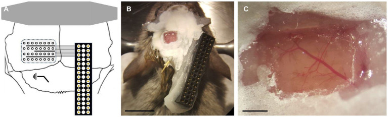
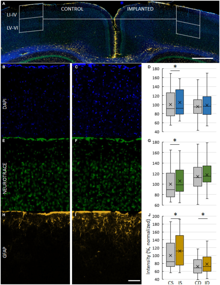
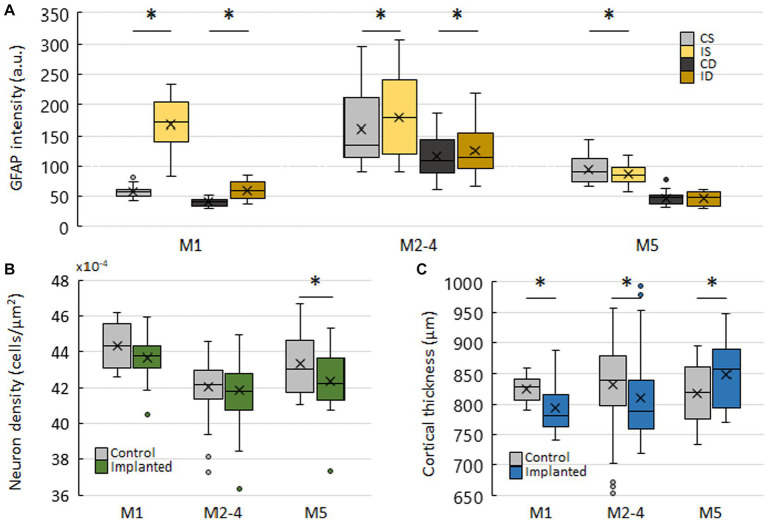
Transparent epidural devices that facilitate the concurrent use of electrophysiology and neuroimaging are arising tools for neuroscience. Testing the biocompatibility and evoked immune response of novel implantable devices is essential to lay down the fundamentals of their extensive application. Here we present an immunohistochemical evaluation of a Parylene HT/indium-tin oxide (ITO) based electrocorticography (ECoG) device, and provide long-term biocompatibility data at three chronic implantation lengths. We implanted Parylene HT/ITO ECoG devices epidurally in 5 mice and evaluated the evoked astroglial response, neuronal density and cortical thickness. We found increased astroglial response in the superficial cortical layers of all mice compared to contralateral unimplanted controls. This difference was largest at the first time point and decreased over time. Neuronal density was lower on the implanted side only at the last time point, while cortical thickness was smaller in the first and second time points, but not at the last. In this study, we present data that confirms the feasibility and chronic use of Parylene HT/ITO ECoG devices.
Keywords: foreign body response; immunohostochemistry; microECoG; parylene; polymer implants.
Copyright © 2023 Madarász, Fedor, Fekete and Rózsa.
Conflict of interest statement
BR was employed by Femtonics Ltd. The remaining authors declare that the research was conducted in the absence of any commercial or financial relationships that could be construed as a potential conflict of interest.
Figures
Similar articles
-
Transparent, low-autofluorescence microECoG device for simultaneous Ca2+ imaging and cortical electrophysiology in vivo.J Neural Eng. 2020 Feb 18;17(1):016062. doi: 10.1088/1741-2552/ab603f. J Neural Eng. 2020. PMID: 31822640
-
Progress in the Field of Micro-Electrocorticography.Micromachines (Basel). 2019 Jan 17;10(1):62. doi: 10.3390/mi10010062. Micromachines (Basel). 2019. PMID: 30658503 Free PMC article. Review.
-
Evaluating the in vivo glial response to miniaturized parylene cortical probes coated with an ultra-fast degrading polymer to aid insertion.J Neural Eng. 2018 Jun;15(3):036002. doi: 10.1088/1741-2552/aa9fad. Epub 2018 Feb 27. J Neural Eng. 2018. PMID: 29485103 Free PMC article.
-
Neuronal excitability and network formation on optically transparent electrode materials.Int IEEE EMBS Conf Neural Eng. 2017 May;2017:154-157. doi: 10.1109/NER.2017.8008315. Epub 2017 Aug 15. Int IEEE EMBS Conf Neural Eng. 2017. PMID: 30338028 Free PMC article.
-
Materials and devices for high-density, high-throughput micro-electrocorticography arrays.Fundam Res. 2024 Feb 28;5(1):17-28. doi: 10.1016/j.fmre.2024.01.016. eCollection 2025 Jan. Fundam Res. 2024. PMID: 40166099 Free PMC article. Review.
Cited by
-
Coniferaldehyde reverses 3-nitropropionic acid-induced Huntington's disease pathologies via PKM2 restoration and JAK2/STAT3 inhibition.Mol Med. 2025 Jul 31;31(1):271. doi: 10.1186/s10020-025-01308-0. Mol Med. 2025. PMID: 40739177 Free PMC article.
-
Hippocampal recording with a soft microelectrode array in a cranial window imaging scheme: a validation study.Sci Rep. 2024 Oct 19;14(1):24585. doi: 10.1038/s41598-024-75170-1. Sci Rep. 2024. PMID: 39427030 Free PMC article.
-
Designing parylene coating for implantable brain-machine interfaces.RSC Adv. 2025 Jul 28;15(33):26660-26672. doi: 10.1039/d5ra04683a. eCollection 2025 Jul 25. RSC Adv. 2025. PMID: 40727297 Free PMC article.
References
LinkOut - more resources
Full Text Sources