

Sensory eye dominance plasticity in the human adult visual cortex
- PMID: 37746154
- PMCID: PMC10513037
- DOI: 10.3389/fnins.2023.1250493
Sensory eye dominance plasticity in the human adult visual cortex
Abstract
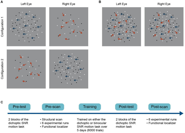
Sensory eye dominance occurs when the visual cortex weighs one eye's data more heavily than those of the other. Encouragingly, mechanisms underlying sensory eye dominance in human adults retain a certain degree of plasticity. Notably, perceptual training using dichoptically presented motion signal-noise stimuli has been shown to elicit changes in sensory eye dominance both in visually impaired and normal observers. However, the neural mechanisms underlying these learning-driven improvements are not well understood. Here, we measured changes in fMRI responses before and after a five-day visual training protocol to determine the neuroplastic changes along the visual cascade. Fifty visually normal observers received training on a dichoptic or binocular variant of a signal-in-noise (left-right) motion discrimination task over five consecutive days. We show significant shifts in sensory eye dominance following training, but only for those who received dichoptic training. Pattern analysis of fMRI responses revealed that responses of V1 and hMT+ predicted sensory eye dominance for both groups, but only before training. After dichoptic (but not binocular) visual training, responses of V1 changed significantly, and were no longer able to predict sensory eye dominance. Our data suggest that perceptual training-driven changes in eye dominance are driven by a reweighting of the two eyes' data in the primary visual cortex. These findings may provide insight into developing region-targeted rehabilitative paradigms for the visually impaired, particularly those with severe binocular imbalance.
Keywords: dichoptic perceptual training; fMRI; perceptual learning; plasticity; sensory eye dominance.
Copyright © 2023 Kam and Chang.
Conflict of interest statement
The authors declare that the research was conducted in the absence of any commercial or financial relationships that could be construed as a potential conflict of interest.
Figures


Similar articles
-
The neurochemistry of learning-driven sensory eye dominance plasticity.Imaging Neurosci (Camb). 2024 Jul 22;2:imag-2-00237. doi: 10.1162/imag_a_00237. eCollection 2024. Imaging Neurosci (Camb). 2024. PMID: 40800464 Free PMC article.
-
Dichoptic Perceptual Training and Sensory Eye Dominance Plasticity in Normal Vision.Invest Ophthalmol Vis Sci. 2021 Jun 1;62(7):12. doi: 10.1167/iovs.62.7.12. Invest Ophthalmol Vis Sci. 2021. PMID: 34106211 Free PMC article. Clinical Trial.
-
Altered Balance of Receptive Field Excitation and Suppression in Visual Cortex of Amblyopic Macaque Monkeys.J Neurosci. 2017 Aug 23;37(34):8216-8226. doi: 10.1523/JNEUROSCI.0449-17.2017. Epub 2017 Jul 25. J Neurosci. 2017. PMID: 28743725 Free PMC article.
-
Real-time modulation of perceptual eye dominance in humans.Proc Biol Sci. 2014 Nov 22;281(1795):20141717. doi: 10.1098/rspb.2014.1717. Proc Biol Sci. 2014. PMID: 25274364 Free PMC article.
-
Sensory Eye Dominance: Relationship Between Eye and Brain.Eye Brain. 2020 Jan 20;12:25-31. doi: 10.2147/EB.S176931. eCollection 2020. Eye Brain. 2020. PMID: 32021530 Free PMC article. Review.
Cited by
-
Effects of binocularity and eye dominance on visually-driven ocular tracking.Front Neurosci. 2025 May 1;19:1504628. doi: 10.3389/fnins.2025.1504628. eCollection 2025. Front Neurosci. 2025. PMID: 40376610 Free PMC article.
-
Improvement of BCI performance with bimodal SSMVEPs: enhancing response intensity and reducing fatigue.Front Neurosci. 2025 Mar 6;19:1506104. doi: 10.3389/fnins.2025.1506104. eCollection 2025. Front Neurosci. 2025. PMID: 40115888 Free PMC article.
-
Interim safety and efficacy of gene therapy for RLBP1-associated retinal dystrophy: a phase 1/2 trial.Nat Commun. 2024 Sep 10;15(1):7438. doi: 10.1038/s41467-024-51575-4. Nat Commun. 2024. PMID: 39256350 Free PMC article. Clinical Trial.
-
The neurochemistry of learning-driven sensory eye dominance plasticity.Imaging Neurosci (Camb). 2024 Jul 22;2:imag-2-00237. doi: 10.1162/imag_a_00237. eCollection 2024. Imaging Neurosci (Camb). 2024. PMID: 40800464 Free PMC article.
References
-
- Beckers G., Hömberg V. (1992). Cerebral visual motion blindness: transitory akinetopsia induced by transcranial magnetic stimulation of human area V5. Proc. R. Soc. Lond. Ser. B Biol. Sci. 249, 173–178. - PubMed
LinkOut - more resources
Full Text Sources