. 2022 Jun 13:3:918717.
doi: 10.3389/ffunb.2022.918717. eCollection 2022.

Antifungal Exposure and Resistance Development: Defining Minimal Selective Antifungal Concentrations and Testing Methodologies

Affiliations

Antifungal Exposure and Resistance Development: Defining Minimal Selective Antifungal Concentrations and Testing Methodologies

Emily M Stevenson et al. Front Fungal Biol. .

Abstract

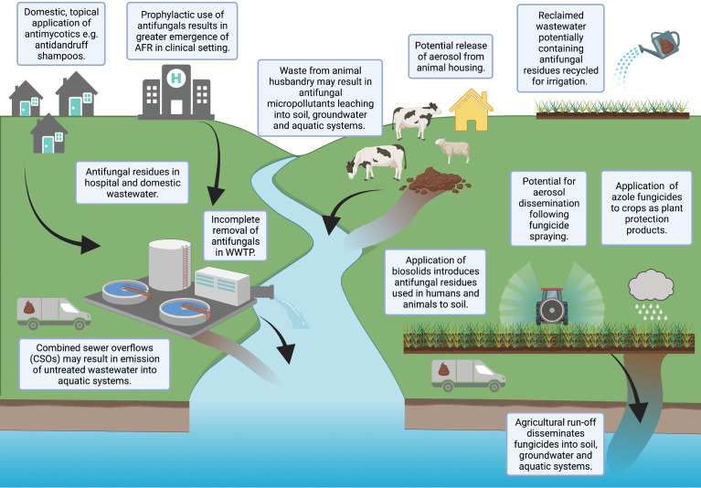
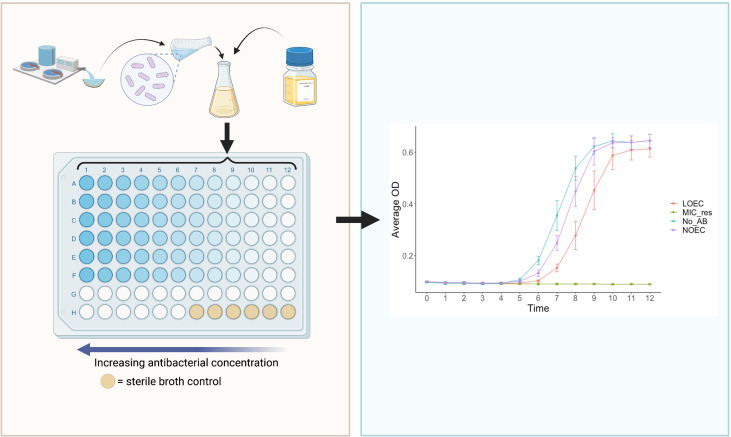
This scoping review aims to summarise the current understanding of selection for antifungal resistance (AFR) and to compare and contrast this with selection for antibacterial resistance, which has received more research attention. AFR is an emerging global threat to human health, associated with high mortality rates, absence of effective surveillance systems and with few alternative treatment options available. Clinical AFR is well documented, with additional settings increasingly being recognised to play a role in the evolution and spread of AFR. The environment, for example, harbours diverse fungal communities that are regularly exposed to antifungal micropollutants, potentially increasing AFR selection risk. The direct application of effect concentrations of azole fungicides to agricultural crops and the incomplete removal of pharmaceutical antifungals in wastewater treatment systems are of particular concern. Currently, environmental risk assessment (ERA) guidelines do not require assessment of antifungal agents in terms of their ability to drive AFR development, and there are no established experimental tools to determine antifungal selective concentrations. Without data to interpret the selective risk of antifungals, our ability to effectively inform safe environmental thresholds is severely limited. In this review, potential methods to generate antifungal selective concentration data are proposed, informed by approaches used to determine antibacterial minimal selective concentrations. Such data can be considered in the development of regulatory guidelines that aim to reduce selection for AFR.

Keywords: antifungal resistance; antifungals; antimicrobial resistance; experimental evolution; fungi; minimal selective concentration; selection.


Conflict of interest statement

The authors declare that the research was conducted in the absence of any commercial or financial relationships that could be construed as a potential conflict of interest.

Figures

Figure 1
Figure 1
Potential pathways for antifungals to enter the environment. Created with BioRender.com.
Figure 2
Figure 2
Schematic diagrams illustrating the MoAs of the primary antifungal drug classes in a fungal cell (A) and primary antibacterial drug classes in a bacterial cell (B). PABA, para-aminobenzoic; DHF, dihydrofolic acid; THF, tetrahydrofolic acid [adapted from Sanseverino et al. (2018)]. Created with BioRender.com.
Figure 3
Figure 3
Schematic diagrams illustrating the known mechanisms of antifungal drug resistance in a fungal cell (A) and antibacterial resistance in a bacterial cell (B). Created with BioRender.com.
Figure 4
Figure 4
Summary of proposed assays to generate antifungal MSC data. Top: serial culture of a fungal community exposed to different antifungal concentrations for a period of 7 days, with daily transfers into fresh nutrient medium and antifungal concentrations. Yellow: phenotypic methods, blue: genotypic methods, purple: methods requiring further investigation. RT-qPCR, reverse transcription quantitative polymerase chain reaction; WGS, whole genome sequencing; PCR, polymerase chain reaction; GC-MS, gas chromatography mass spectroscopy. Created with BioRender.com.
Figure 5
Figure 5
The ‘SELection End points in Communities of bacTeria’ (SELECT) Method (Murray et al., 2020). Left: a schematic overview of the SELECT assay; a 96-well-plate containing Iso-Sensitest broth inoculated with a complex sewage community, exposed to two-fold dilutions of an antibacterial. Right: an expected graphical output of the SELECT method, showing complex bacterial community growth curves in response to different concentrations of antibacterials. The LOEC is identified where the growth of the community is significantly lower than the no-antibacterial control, at the time point which exhibits the strongest dose-response relationship. LOEC, Lowest Observed Effect Concentration; NOEC, No Observed Effect Concentration; MIC_res, minimum inhibitory concentration of resistant strains, No_AB, No Antibacterial control. Created with BioRender.com.

Similar articles

Cited by

References

    1. Anderson J. B. (2005). Evolution of Antifungal-Drug Resistance: Mechanisms and Pathogen Fitness. Nat. Rev. Microbiol. 3 (7), 547–556. doi: 10.1038/nrmicro1179 - DOI - PubMed
    1. Andersson D. I., Hughes D. (2010). Antibiotic Resistance and Its Cost: Is it Possible to Reverse Resistance? Nat. Rev. Microbiol. 8 (4), 260–271. doi: 10.1038/nrmicro2319 - DOI - PubMed
    1. Andersson D. I., Hughes D. (2012). Evolution of Antibiotic Resistance at Non-Lethal Drug Concentrations. Drug Resist. Update 15 (3), 162–172. doi: 10.1016/j.drup.2012.03.005 - DOI - PubMed
    1. Arai T., Majima H., Watanabe A., Kamei K. (2020). A Simple Method To Detect Point Mutations in Aspergillus Fumigatus Cyp51a Gene Using a Surveyor Nuclease Assay. Antimicrob. Agents Chemother. 64 (4), e02271–19. doi: 10.1128/AAC.02271-19 - DOI - PMC - PubMed
    1. Arendrup M. C., Meletiadis J., Mouton J. W., Guinea J., Cuenca-Estrella M., Lagrou K., et al. . (2016). EUCAST Technical Note on Isavuconazole Breakpoints for Aspergillus, Itraconazole Breakpoints for Candida and Updates for the Antifungal Susceptibility Testing Method Documents. Clin. Microbiol. Infect. 22 (6), 571 e571–574. doi: 10.1016/j.cmi.2016.01.017 - DOI - PubMed

LinkOut - more resources