. 2023 Jun 12:30:90-102.
doi: 10.1016/j.omtm.2023.06.002. eCollection 2023 Sep 14.

Innate and adaptive AAV-mediated immune responses in a mouse model of Duchenne muscular dystrophy

Affiliations

Innate and adaptive AAV-mediated immune responses in a mouse model of Duchenne muscular dystrophy

Michael R Emami et al. Mol Ther Methods Clin Dev. .

Abstract

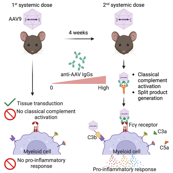
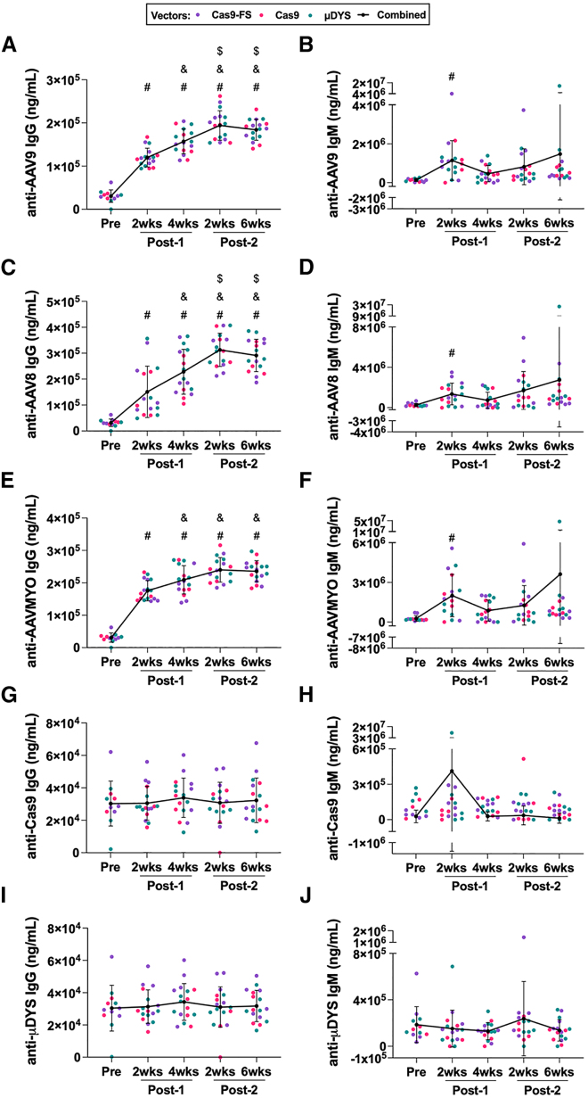
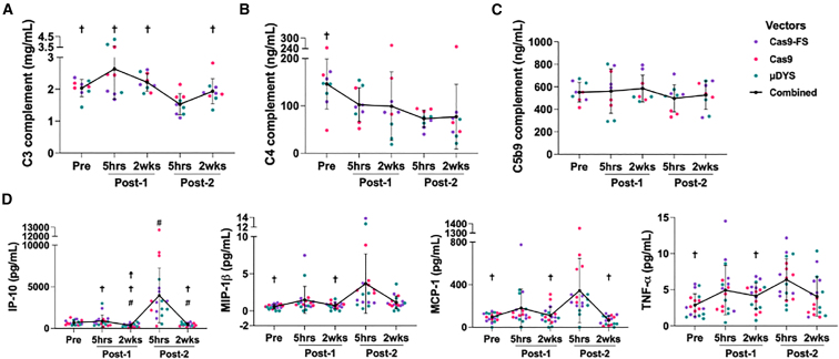
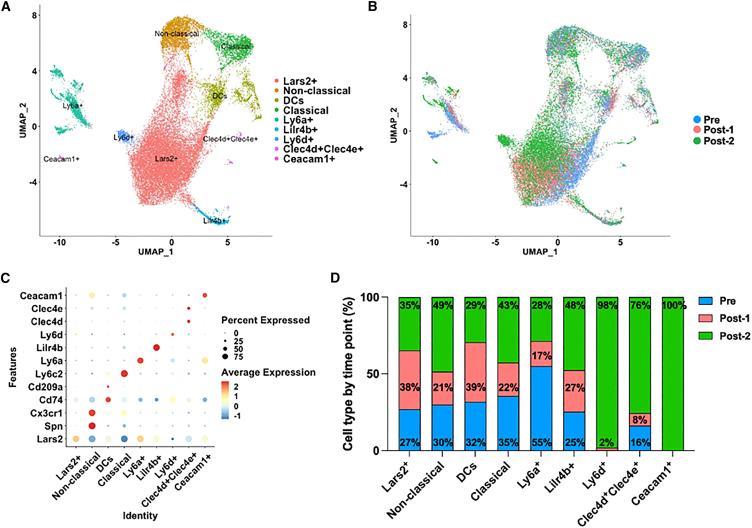
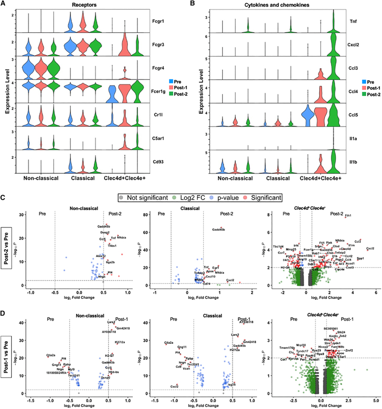
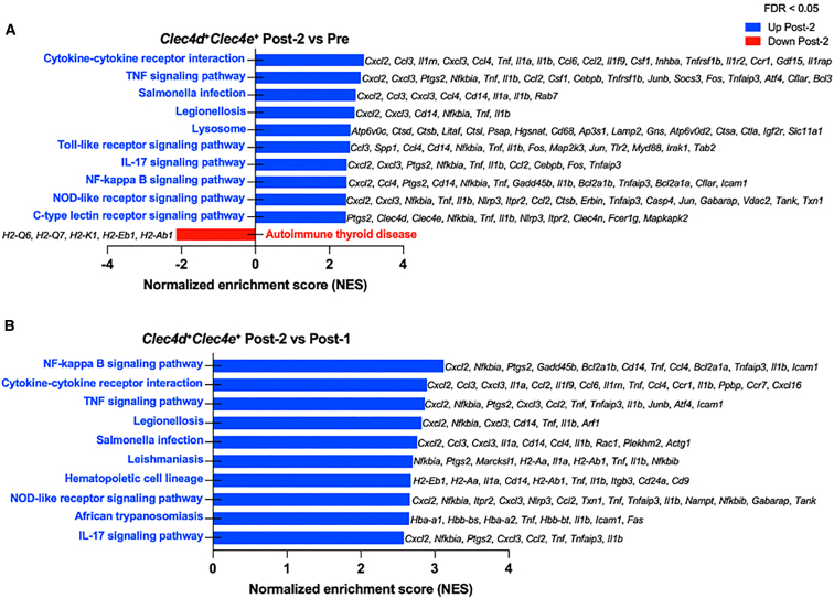
High systemic doses of adeno-associated viruses (AAVs) have been associated with immune-related serious adverse events (SAEs). Although AAV was well tolerated in preclinical models, SAEs were observed in clinical trials, indicating the need for improved preclinical models to understand AAV-induced immune responses. Here, we show that mice dual-dosed with AAV9 at 4-week intervals better recapitulate aspects of human immunity to AAV. In the model, anti-AAV9 immunoglobulin G (IgGs) increased in a linear fashion between the first and second AAV administrations. Complement activation was only observed in the presence of high levels of both AAV and anti-AAV IgG. Myeloid-derived pro-inflammatory cytokines were significantly induced in the same pattern as complement activation, suggesting that myeloid cell activation to AAV may rely on the presence of both AAV and anti-AAV IgG complexes. Single-cell RNA sequencing of peripheral blood mononuclear cells confirmed that activated monocytes were a primary source of pro-inflammatory cytokines and chemokines, which were significantly increased after a second AAV9 exposure. The same activated monocyte clusters expressed both Fcγ and complement receptors, suggesting that anti-AAV-mediated activation of myeloid cells through Fcγ receptors and/or complement receptors is one mechanism by which anti-AAV antigen complexes may prime antigen-presenting cells and amplify downstream immunity.

Keywords: Luminex; adaptive immunity; adeno-associated virus; innate immunity; muscular dystrophy; single-cell RNA sequencing.


Conflict of interest statement

M.J.S., A.D.P., and C.S.Y. are co-founders of MyoGene Bio, a startup spun out of UCLA developing gene editing therapies for Duchenne muscular dystrophy.

Figures

None
Graphical abstract
Figure 1
Figure 1
Assessment of anti-AAV and anti-transgene antibody responses using a high-content protein microarray (HCPM) (A and B) Anti-AAV9 IgG and IgM responses were measured via HCPM from male (n = 9) and female (n = 8) mdx mice at indicated time points. The key indicates the vector that was dosed and the black line represents AAV9-treated groups combined by time point, which was used for multiple comparison statistical analysis. Error bars for all HCPM graphs represent standard deviation. Symbols above time points are used to represent statistical significance, in which p < 0.05, for time points compared with: (#) Pre, (&) Post-1 (2 weeks), and ($) Post-1 (4 weeks). The data and levels of statistical significance between time point comparisons are available in Tables S1 and S2. (C–F) Plasma collected from mice double-dosed with AAV9 was assessed by HCPM for the ability to bind to other AAV serotypes. Capsids of AAV8 and AAVMYO were printed on the chip and plasma from male (n = 9) and female (n = 8) mdx mice was assessed. (G–J) anti-Cas9 and anti-μDYS IgG and IgM responses were assayed via HCPM from male (n = 9) and female (n = 8) mdx mice at indicated time points.
Figure 2
Figure 2
Consumption of complement components, C3 and C4, and concomitant induction of pro-inflammatory chemokines and cytokines after the second dose of AAV (A–C) Plasma from mdx mice was evaluated by ELISA for levels of complement C3 (n = 9 females), C4 (n = 9 females), and C5b9 (n = 9 females). C3 levels for males (n = 9) and females (n = 9) are shown in Figure S3A. The key indicates the vector that was dosed and the black line represents AAV9-treated groups combined by time point, which was used for multiple comparison statistical analysis. Error bars for all graphs represent standard deviation. Symbols above time points are used to represent statistical significance, in which p < 0.05, for time points compared with (†) Post-2 (5 h). The data and levels of statistical significance between time point comparisons are available in Tables S3 and S4. (D) Levels of immunomodulatory analytes: IP-10 (CXCL10), MIP-1β (CCL4), MCP-1 (CCL2), and TNF-α as measured by Luminex ProcartaPlex, n = 9 males and n = 9 females. Symbols above time points are used to represent statistical significance, in which p < 0.05, for time points compared with (#) Pre, (☨) Post-1 (5 h), and (†) Post-2 (5 h). The data and levels of statistical significance between time point comparisons are available in Tables S5 and S6.
Figure 3
Figure 3
Unbiased characterization via scRNA-seq analysis to identify cell types responding to AAV in peripheral blood mononuclear cells (PBMCs) (A) Monocyte subcluster UMAP shows heterogeneous sub-populations present in AAV9 double-dosed mice. (B) Monocyte subcluster UMAP, color coded by time point before and after AAV dosing. Pre (blue), Post-1 (pink) and Post-2 (green). (C) Dot plot of top differentially expressed genes among monocyte sub-populations. Color is scaled by average expression and the dot size is proportional to the percent of cells expressing the respective gene. (D) Bar graph shows the percentage of monocyte sub-populations by time point. Pre (blue), Post-1 (pink) and Post-2 (green).
Figure 4
Figure 4
Monocyte sub-populations demonstrate activation and induction of pro-inflammatory chemokine and cytokine genes after the second, not the first, systemic administration of AAV9 (A) Stacked violin plot shows expression of FcγRs and complement receptor genes present in activated non-classical, classical, and Clec4d+Clec4e+ monocyte populations. (B) Stacked violin plot shows the expression of significantly differentially expressed pro-inflammatory cytokine and chemokine genes upregulated Post-2 (2 weeks). (C and D) Volcano plots of classical, non-classical, and Clec4d+Clec4e+ monocytes showing differentially expressed genes between Post-2 (2 weeks) and Pre (C) and Post-1 (2 weeks) and Pre (D). Significantly up- and down-regulated genes contain a Log2(Fold Change) > 0.5 or Log2(Fold Change) < 0.5 and -Log10(p value) > 2.0.
Figure 5
Figure 5
Gene sent enrichment analysis of Clec4d+Clec4e+ monocytes reveal inflammatory response pathways enriched after AAV re-administration (A) Differential gene expression analysis between Post-2 and Pre time points and gene set enrichment analysis (GSEA) identified inflammatory response pathways including cytokine signaling, TLR signaling, and NF-κB signaling. Table of GSEA pathways and associated genes are listed in Table S7. (B) Differential gene expression analysis between Post-2 and Post-1 time points and GSEA similarly identified inflammatory response pathways enriched after AAV re-administration compared with the first administration. Table of GSEA pathways and associated genes are listed in Table S8.

References

    1. Greer C., Kozyak B., Stedman H. Challenges at the Crossroads: Myopathy Trials in 2020 Hindsight. Mol. Ther. 2021;29:420–421. doi: 10.1016/j.ymthe.2021.01.012. - DOI - PMC - PubMed
    1. Manini A., Abati E., Nuredini A., Corti S., Comi G.P. Adeno-Associated Virus (AAV)-Mediated Gene Therapy for Duchenne Muscular Dystrophy: The Issue of Transgene Persistence. Front. Neurol. 2021;12:814174. doi: 10.3389/fneur.2021.814174. - DOI - PMC - PubMed
    1. Mendell J.R., Al-Zaidy S.A., Rodino-Klapac L.R., Goodspeed K., Gray S.J., Kay C.N., Boye S.L., Boye S.E., George L.A., Salabarria S., et al. Current Clinical Applications of In Vivo Gene Therapy with AAVs. Mol. Ther. 2021;29:464–488. doi: 10.1016/j.ymthe.2020.12.007. - DOI - PMC - PubMed
    1. Fortunato F., Rossi R., Falzarano M.S., Ferlini A. Innovative Therapeutic Approaches for Duchenne Muscular Dystrophy. J. Clin. Med. 2021;10:820. doi: 10.3390/jcm10040820. - DOI - PMC - PubMed
    1. Chand D.H., Zaidman C., Arya K., Millner R., Farrar M.A., Mackie F.E., Goedeker N.L., Dharnidharka V.R., Dandamudi R., Reyna S.P. Thrombotic Microangiopathy Following Onasemnogene Abeparvovec for Spinal Muscular Atrophy: A Case Series. J. Pediatr. 2021;231:265–268. doi: 10.1016/j.jpeds.2020.11.054. - DOI - PubMed