. 2023 May 18:30:30-47.
doi: 10.1016/j.omtm.2023.05.017. eCollection 2023 Sep 14.

Evaluation of an AAV9-mini-dystrophin gene therapy candidate in a rat model of Duchenne muscular dystrophy

Affiliations

Evaluation of an AAV9-mini-dystrophin gene therapy candidate in a rat model of Duchenne muscular dystrophy

Caroline Le Guiner et al. Mol Ther Methods Clin Dev. .

Abstract

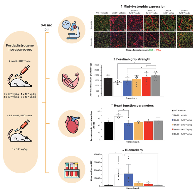
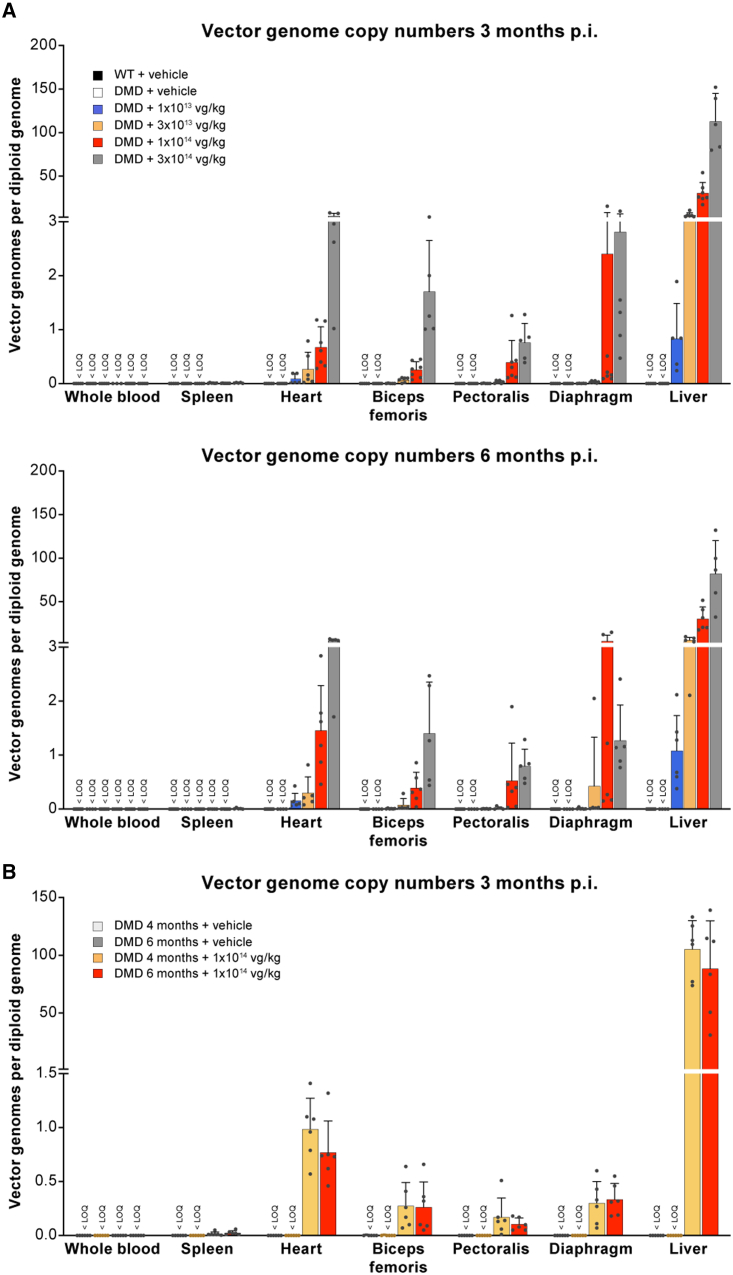
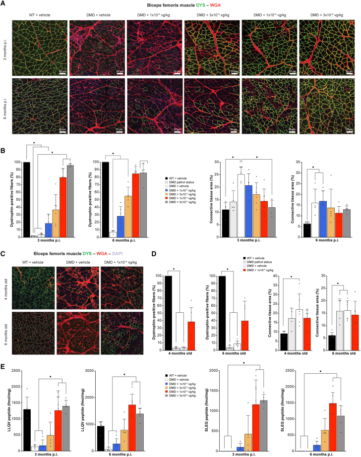
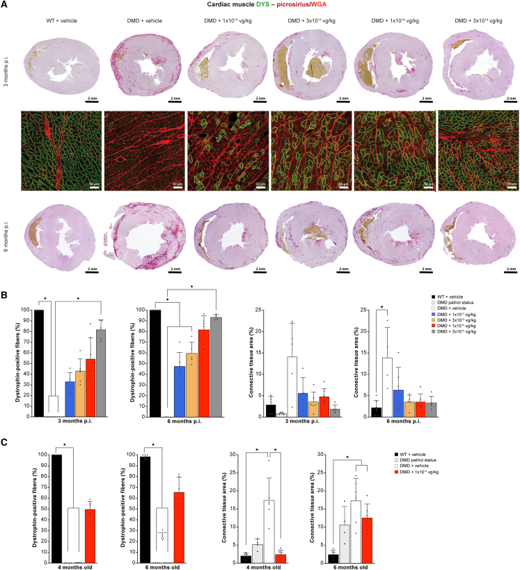
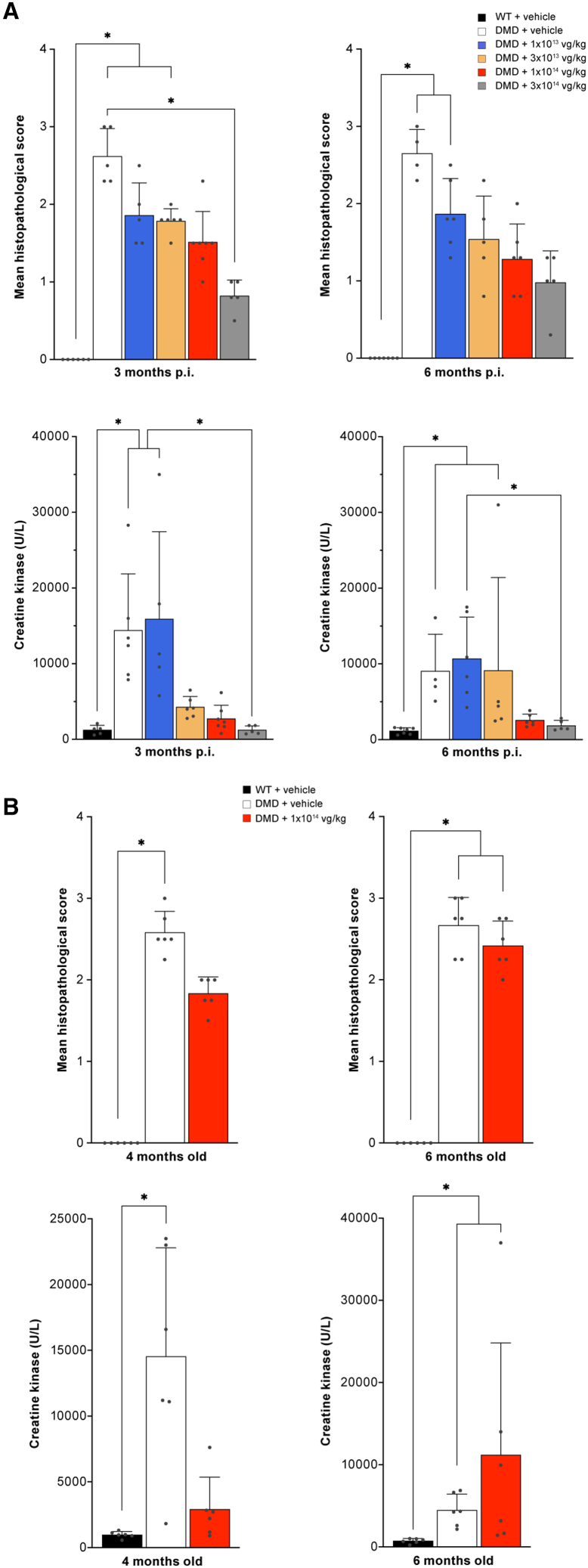
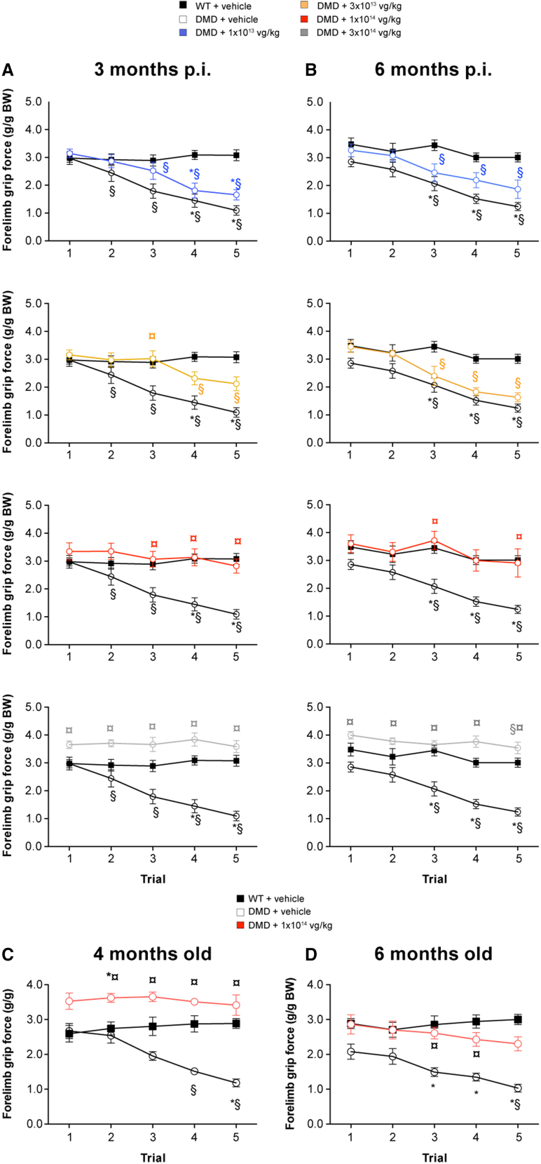
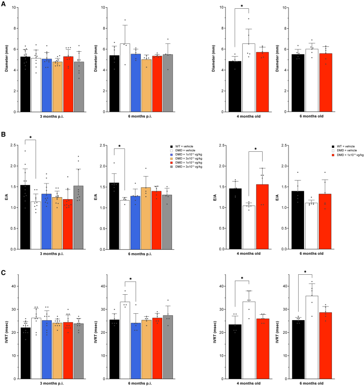
Duchenne muscular dystrophy (DMD) is an X-linked disease caused by loss-of-function mutations in the dystrophin gene and is characterized by muscle wasting and early mortality. Adeno-associated virus-mediated gene therapy is being investigated as a treatment for DMD. In the nonclinical study documented here, we determined the effective dose of fordadistrogene movaparvovec, a clinical candidate adeno-associated virus serotype 9 vector carrying a human mini-dystrophin transgene, after single intravenous injection in a dystrophin-deficient (DMDmdx) rat model of DMD. Overall, we found that transduction efficiency, number of muscle fibers expressing the human mini-dystrophin polypeptide, improvement of the skeletal and cardiac muscle tissue architecture, correction of muscle strength and fatigability, and improvement of diastolic and systolic cardiac function were directly correlated with the amount of vector administered. The effective dose was then tested in older DMDmdx rats with a more dystrophic phenotype similar to the pathology observed in older patients with DMD. Except for a less complete rescue of muscle function in the oldest cohort, fordadistrogene movaparvovec was also found to be therapeutically effective in older DMDmdx rats, suggesting that this product may be appropriate for evaluation in patients with DMD at all stages of disease.

Keywords: DMDmdx rat; Duchenne muscular dystrophy; age; dose study; gene therapy; mini-dystrophin; recombinant adeno-associated virus.


Figures

None
Graphical abstract
Figure 1
Figure 1
Vector copy numbers per diploid genomes in whole blood and tissue samples from DMDmdx rats (A) Data from DMDmdx rats injected at 2 months of age and sacrificed at 3 (top) or 6 (bottom) months p.i. (B) Data from DMDmdx rats injected at 4 or 6 months of age and sacrificed at 3 months p.i. Vector copy numbers per diploid genomes were determined using qPCR. Results are expressed as the mean ± SEM. Limit of quantification = 0.002 vg/dg. DMDmdx, dystrophin-deficient rat; p.i., post injection; qPCR, quantitative polymerase chain reaction; SEM, standard error of the mean.
Figure 2
Figure 2
Immunohistochemical analysis of mini-dystrophin expression and assessment of fibrosis in the biceps femoris muscle (A) Representative images of biceps femoris muscle sections collected 3 and 6 months p.i. from the following groups of rats 2 months old at the time of injection: WT+vehicle, DMDmdx pathol status, DMDmdx+vehicle and vector-injected DMDmdx rats. The NCL-DYSB antibody was used to detect dystrophin/mini-dystrophin expression (green), and Alexa Fluor 555 WGA conjugate was used to detect connective tissue (red). DRAQ5 fluorescent DNA dye was used to detect nuclei. (B) Quantification of dystrophin and fibrosis labeling in sections from 2-month-old treated rats (samples collected at 3 or 6 months p.i.). Left panel: percentage of dystrophin-positive fibers. Right panel: percent area occupied by WGA-labeled connective tissue. (C) Representative images of biceps femoris muscle sections collected 3 months p.i. from the following groups of rats 4 and 6 months old at the time of injection (samples collected at 3 months p.i.): WT+vehicle-, DMDmdx+vehicle-, and vector-injected DMDmdx rats. The NCL-DYSB antibody was used to detect dystrophin/mini-dystrophin expression (green), and Alexa Fluor 555 WGA conjugate was used to detect connective tissue (red). DRAQ5 fluorescent DNA dye was used to detect nuclei. (D) Quantification of dystrophin and fibrosis labeling in sections from 4- to 6-month-old treated rats (samples collected at 3 months p.i.). Left panel: percentage of dystrophin-positive fibers (mean ± SD). Right panel: percent area occupied by connective tissue as labeled by WGA (mean ± SD). (E) IA-LC-MS/MS-based quantification of dystrophin/mini-dystrophin expression levels in biceps femoris samples from 2-month-old treated rats (samples collected at 3 or 6 months p.i.). Left panel: total dystrophin expression levels based on peptide LLQV. Right panel: mini-dystrophin expression levels based on peptide SLEG. Results are expressed as the mean ± SEM. ∗p < 0.05 (nonparametric Kruskal-Wallis test followed by a post hoc Dunn’s multiple-comparisons test). WGA, wheat germ agglutinin; WT, wild-type; pathol, pathological.
Figure 3
Figure 3
Immunohistochemical analysis of mini-dystrophin expression and assessment of fibrosis in the heart (A) Transverse sections done at the level of the apex were stained NCL-DYSB antibody to detect dystrophin/mini-dystrophin expression (green), with picrosirius red for connective tissue (purple) and WGA conjugate (red) for connective tissue. Representative images correspond to samples collected 3 and 6 months p.i., from WT+vehicle, DMDmdx+vehicle, and vector-injected DMDmdx rats. (B) Quantification of dystrophin and fibrosis labeling in sections from 2-month-old treated rats (samples collected at 3 or 6 months p.i.). Left panel: percentage of dystrophin-positive fibers. Right panel: percent area occupied by picrosirius red-positive connective tissue. (C) Quantification of dystrophin and fibrosis labeling in sections from 4- and 6-month-old treated rats (samples collected 3 months p.i.). Left panel: percentage of dystrophin-positive fibers. Right panel: percent area occupied by picrosirius red-positive connective tissue. Results are expressed as the mean ± SEM. ∗p < 0.05 (nonparametric Kruskal-Wallis test, followed by a post hoc Dunn’s multiple-comparisons test).
Figure 4
Figure 4
Muscle histopathology and serum CK levels (A) Muscle histopathological score obtained after histopathological observation of muscle samples obtained at 3 and 6 months p.i. in rats injected at 2 months of age and serum CK levels (U/L) obtained in the same animals. (B) Muscle histopathological score obtained after histopathological observation of muscle samples obtained at 3 months p.i. in rats injected at 4 and 6 months of age and serum CK levels (U/L) obtained in the same animals. Results are expressed as the mean ± SEM. ∗p < 0.05 (nonparametric Kruskal-Wallis test followed by a post hoc Dunn’s multiple-comparisons test).
Figure 5
Figure 5
Assessment of mini-dystrophin expression on skeletal muscle function (A and B) Dose-range study of vector injected in DMDmdx rats at 2 months of age and evaluated in grip strength assessments over 5 successive trials (grip strength tests) at 3 (A) and 6 (B) months p.i. Data plots show the mean forelimb grip force (g/g normalized to body weight [BW]) ± SEM collected at each trial for each dose of vector. (C) Evaluation of muscle function in DMDmdx rats injected with 1 × 1014 vg/kg vector at 4 and 6 months of age (grip strength tests, as described above). Data represent the mean forelimb grip force (g/g normalized to BW) ± SEM collected at each trial 3 months p.i. Left panel: rats dosed at 4 months of age. Right panel: rats dosed at 6 months of age. ∗p < 0.05 vs. WT. ¤p < 0.05 vs. DMDmdx control rats. §p < 0.05 vs. trial 1 (non-parametric Friedman test followed by Dunn’s post hoc test).
Figure 6
Figure 6
Assessment of mini-dystrophin expression on cardiac muscle function (A) LV diastolic diameter measured during diastole from long-axis images obtained by M-mode echocardiogram. (B) E/A ratio measured using pulsed Doppler with an apical 4-chamber orientation. (C) IVRT measured using pulsed Doppler with apical 4-chamber orientation. Results are expressed as the mean ± SEM. ∗p < 0.05 (nonparametric Kruskal-Wallis test followed by a post-hoc Dunn’s multiple-comparisons test). E/A, early/late diastolic velocity; IVRT, isovolumetric relaxation time; LV, left ventricle.

Similar articles

Cited by

References

    1. Hoffman E.P., Brown R.H., Jr., Kunkel L.M. Dystrophin: the protein product of the Duchenne muscular dystrophy locus. Cell. 1987;51:919–928. doi: 10.1016/0092-8674(87)90579-4. - DOI - PubMed
    1. Mendell J.R., Lloyd-Puryear M. Report of MDA muscle disease symposium on newborn screening for Duchenne muscular dystrophy. Muscle Nerve. 2013;48:21–26. doi: 10.1002/mus.23810. - DOI - PubMed
    1. Michele D.E., Campbell K.P. Dystrophin-glycoprotein complex: post-translational processing and dystroglycan function. J. Biol. Chem. 2003;278:15457–15460. doi: 10.1074/jbc.R200031200. - DOI - PubMed
    1. Bogdanovich S., Krag T.O.B., Barton E.R., Morris L.D., Whittemore L.A., Ahima R.S., Khurana T.S. Functional improvement of dystrophic muscle by myostatin blockade. Nature. 2002;420:418–421. doi: 10.1038/nature01154. - DOI - PubMed
    1. Boland B.J., Silbert P.L., Groover R.V., Wollan P.C., Silverstein M.D. Skeletal, cardiac, and smooth muscle failure in Duchenne muscular dystrophy. Pediatr. Neurol. 1996;14:7–12. doi: 10.1016/0887-8994(95)00251-0. - DOI - PubMed

LinkOut - more resources