. 2023 Jun 25:30:181-190.
doi: 10.1016/j.omtm.2023.06.008. eCollection 2023 Sep 14.

Systemic gene therapy using an AAV44.9 vector rescues a neonatal lethal mouse model of propionic acidemia

Affiliations

Systemic gene therapy using an AAV44.9 vector rescues a neonatal lethal mouse model of propionic acidemia

Randy J Chandler et al. Mol Ther Methods Clin Dev. .

Abstract

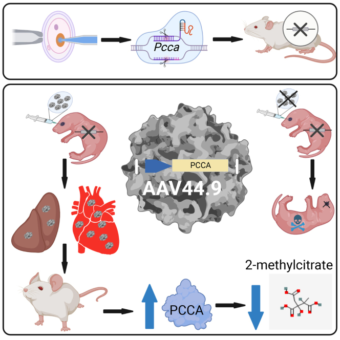
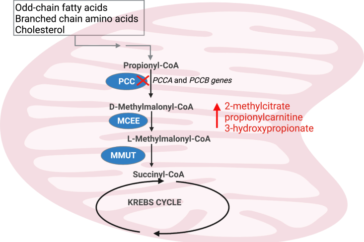
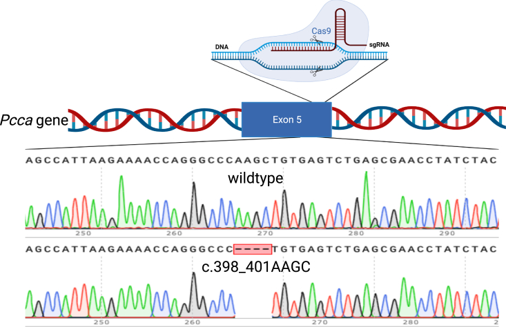
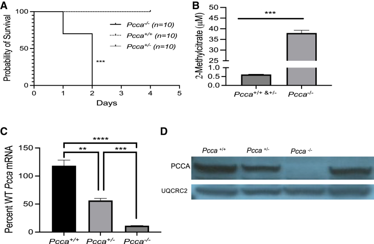
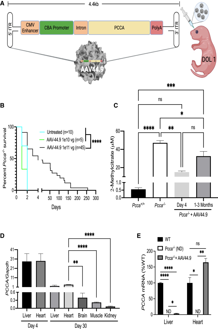
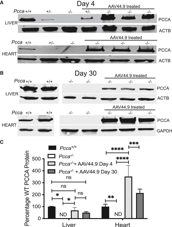
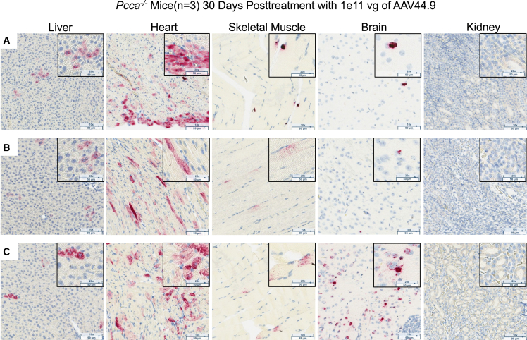
Propionic acidemia (PA) is rare autosomal recessive metabolic disorder caused by defects in the mitochondrially localized enzyme propionyl-coenzyme A (CoA) carboxylase. Patients with PA can suffer from lethal metabolic decompensation and cardiomyopathy despite current medical management, which has led to the pursuit of gene therapy as a new treatment option for patients. Here we assess the therapeutic efficacy of a recently described adeno-associated virus (AAV) capsid, AAV44.9, to deliver a therapeutic PCCA transgene in a new mouse model of propionyl-CoA carboxylase α (PCCA) deficiency generated by genome editing. Pcca-/- mice recapitulate the severe neonatal presentation of PA and manifest uniform neonatal lethality, absent PCCA expression, and increased 2-methylcitrate. A single injection of the AAV44.9 PCCA vector in the immediate newborn period, systemically delivered at a dose of 1e11 vector genome (vg)/pup but not 1e10 vg/pup, increased survival, reduced plasma methylcitrate, and resulted in high levels of transgene expression in the liver and heart in treated Pcca-/- mice. Our studies not only establish a versatile and accurate new mouse model of PA but further demonstrate that the AAV44.9 vectors may be suitable for treatment of many metabolic disorders where hepato-cardiac transduction following systemic delivery is desired, such as PA, and, by extension, fatty acid oxidation defects and glycogen storage disorders.

Keywords: AAV44.9; PCCA; cardiac tropism; genome editing; mouse model; neonatal gene therapy; organic acidemia; propionic acidemia; propionylcoa carboxylase deficiency; systemic gene therapy.


Conflict of interest statement

R.J.C., G.D.P., J.A.C., and C.P.V. are inventors on a patent application filed by the NIH on their behalf on use of AAV44.9 as a gene therapy vector to treat methylmalonic acidemia.

Figures

None
Graphical abstract
Figure 1
Figure 1
Mitochondrial catabolism of propionyl-CoA to succinyl-CoA Propionic acidemia (PA) is caused by mutations in the PCCA or PCCB gene, which encode for the respective subunits of propionyl-CoA carboxylase (PCC). Enzymes downstream of PCC in the metabolism of propionyl-CoA to succinyl-CoA, a Krebs cycle intermediate. Other enzymes include D-methylmalonyl-CoA epimerase (MCEE) and methylmalonyl-CoA mutase (MMUT). The figure was created with BioRender.
Figure 2
Figure 2
Schematic of the Pccac.398_401delAAGC allele Exon 5 of the Pcca was targeted with a single-guide RNA (sgRNA) resulting in a 4-bp deletion after Cas9 cleavage and repair by non-homologous end joining (NEHJ). Shown are Sanger sequencing results of genomic DNA from a mouse with a WT Pcca genotype compared with a mouse homozygous for the Pccac.398_401delAAGC mutation (bottom). The figure was created with BioRender.
Figure 3
Figure 3
Clinical and biochemical characterization of Pcca−/− mice (A) Survival of mice with Pcca+/+,Pcca+/−, and a Pcca−/− genotypes. (B) Plasma 2-methylcitrate levels from Pcca+/+ and Pcca+/− newborn pups (n = 13) in comparison with Pcca−/− newborn pups (n = 6). (C) Pcca mRNA levels in the liver of Pcca+/+ (n = 3), Pcca+/− (n = 3), and Pcca−/− (n = 3) mice, shown as a percentage of WT Pcca mRNA expression normalized to Actb. (D) Immunoblot of 50 μg hepatic protein from Pcca+/+, Pcca+/−, and Pcca−/− newborn pups for the PCCA protein using the mitochondrial protein ubiquinol-cytochrome c reductase core protein 2 (UQCRC2) as a loading control. ∗∗p < 0.01, ∗∗∗p < 0.001, ∗∗∗∗p < 0.0001.
Figure 4
Figure 4
AAV44.9 gene delivery of PCCA rescues Pcca−/− mice from neonatal lethality (A) Schematic of the recombinant AAV vector genome (pAAV-CBA-PCCA) packaged with the AAV44.9 capsid (CMV, cytomegalovirus; CBA, chicken β-actin). Pcca−/− pups received a retro-orbital injection of 1e10 vg or 1e11 vg of AAV44.9-CBA-PCCA on day of life 1 (DOL 1). (B) Survival curve comparing the viability of Pcca−/− mice treated with AAV44.9 at doses of 1e10 vg or 1e11 vg with untreated Pcca−/− mice. (C) Plasma 2-methylcitrate in untreated neonatal Pcca−/− mice and Pcca−/− mice 4 days and 1–3 months after treatment with AAV44.9 at 1e11 vg. (D) Vector biodistribution in liver, heart, brain skeletal muscle, and kidneys from Pcca−/− treated mice (n = 3) 4 days and 1 month post treatment, as determined by ddPCR. (E) PCCA and Pcca mRNA expression in the liver and heart (WT, n = 3, 3; PA, n = 2, 1; AAV44.9, n = 3, 3), shown as a percentage of WT Pcca mRNA expression normalized to Actb (liver) or Gapdh (heart). ∗p < 0.05, ∗∗p < 0.01, ∗∗∗p < 0.001, ∗∗∗∗p < 0.0001; ns, not significant; ND, not detected.
Figure 5
Figure 5
Expression of PCCA protein in the liver and heart (A) Immunoblot of hepatic and cardiac PCCA protein from untreated Pcca+/+ (n = 1), Pcca−/− (n = 1), and Pcca+/− (n = 1) and AAV44.9-treated Pcca+/− (n = 1) and Pcca−/− (n = 3) mice 4 days post treatment. (B) Immunoblot of hepatic and cardiac PCCA protein from untreated Pcca+/+ (n = 2) and Pcca−/− (n = 2) and AAV44.9-treated Pcca−/− mice (n = 3) 30 days post treatment. (C) Quantification of PCCA protein expression relative to untreated Pcca+/+ mice from the blots depicted in (A) and (B). ∗p < 0.05, ∗∗p < 0.003, ∗∗∗p < 0.001, ∗∗∗∗p < 0.0001.
Figure 6
Figure 6
RNA in situ hybridization in tissues 30 days after AAV44.9 gene delivery to detect PCCA expression PCCA transgene mRNA (punctate red staining) in situ hybridization (magnification, 10× with 20× inset) of liver, heart, skeletal muscle, brain, and kidney tissue from three Pcca−/− AAV44.9-CBA-PCCA-treated mice, labeled A, B, and C.

Similar articles

Cited by

References

    1. Almási T., Guey L.T., Lukacs C., Csetneki K., Vokó Z., Zelei T. Systematic literature review and meta-analysis on the epidemiology of propionic acidemia. Orphanet J. Rare Dis. 2019;14:40. doi: 10.1186/s13023-018-0987-z. - DOI - PMC - PubMed
    1. Shchelochkov O.A., Carrillo N., Venditti C. In: GeneReviews((R)) Adam M.P., Everman D.B., Mirzaa G.M., Pagon R.A., Wallace S.E., Bean L.J.H., Gripp K.W., Amemiya A., editors. 1993. Propionic Acidemia.
    1. Gompertz D., Storrs C.N., Bau D.C., Peters T.J., Hughes E.A. Localisation of enzymic defect in propionicacidaemia. Lancet. 1970;1:1140–1143. doi: 10.1016/s0140-6736(70)91216-x. - DOI - PubMed
    1. Hsia Y.E., Scully K.J., Rosenberg L.E. Inherited propionyl-Coa carboxylase deficiency in "ketotic hyperglycinemia". J. Clin. Invest. 1971;50:127–130. doi: 10.1172/JCI106466. - DOI - PMC - PubMed
    1. Huang C.S., Sadre-Bazzaz K., Shen Y., Deng B., Zhou Z.H., Tong L. Crystal structure of the alpha(6)beta(6) holoenzyme of propionyl-coenzyme A carboxylase. Nature. 2010;466:1001–1005. doi: 10.1038/nature09302. - DOI - PMC - PubMed

LinkOut - more resources