

Organoids and metastatic orthotopic mouse model for mismatch repair-deficient colorectal cancer
- PMID: 37746286
- PMCID: PMC10516605
- DOI: 10.3389/fonc.2023.1223915
Organoids and metastatic orthotopic mouse model for mismatch repair-deficient colorectal cancer
Abstract
Background: Genome integrity is essential for the survival of an organism. DNA mismatch repair (MMR) genes (e.g., MLH1, MSH2, MSH6, and PMS2) play a critical role in the DNA damage response pathway for genome integrity maintenance. Germline mutations of MMR genes can lead to Lynch syndrome or constitutional mismatch repair deficiency syndrome, resulting in an increased lifetime risk of developing cancer characterized by high microsatellite instability (MSI-H) and high mutation burden. Although immunotherapy has been approved for MMR-deficient (MMRd) cancer patients, the overall response rate needs to be improved and other management options are needed.
Methods: To better understand the biology of MMRd cancers, elucidate the resistance mechanisms to immune modulation, and develop vaccines and therapeutic testing platforms for this high-risk population, we generated organoids and an orthotopic mouse model from intestine tumors developed in a Msh2-deficient mouse model, and followed with a detailed characterization.
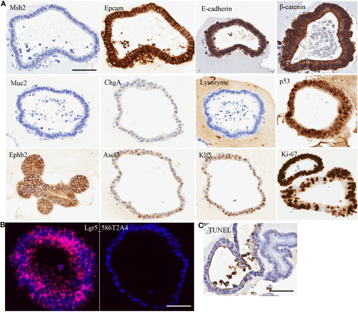
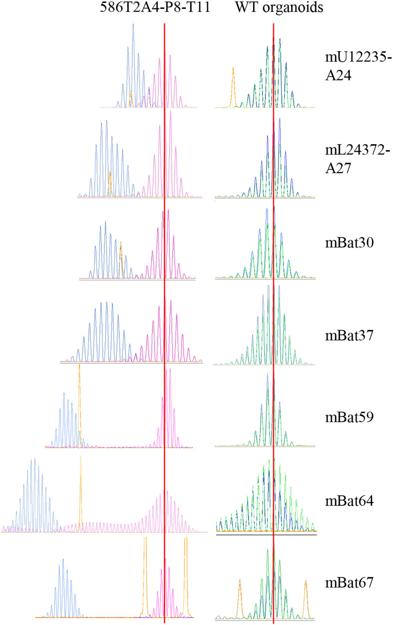
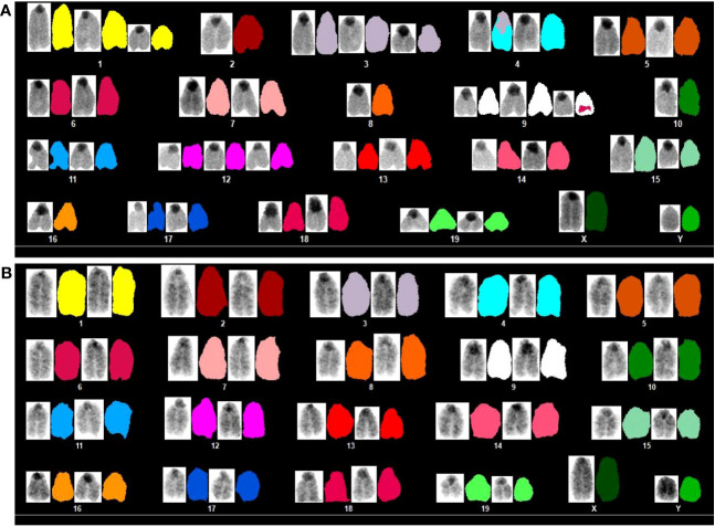
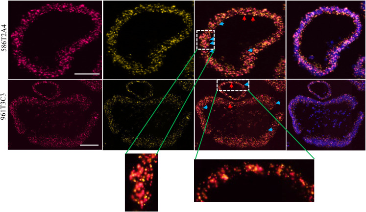
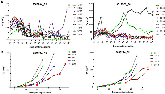
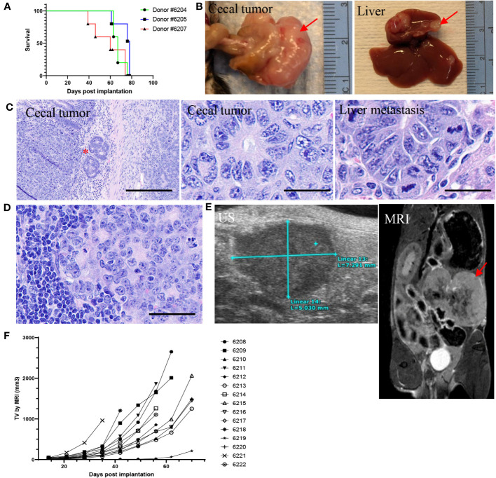
Results: The organoids were shown to be of epithelial origin with stem cell features, to have a high frameshift mutation frequency with MSI-H and chromosome instability, and intra- and inter-tumor heterogeneity. An orthotopic model using intra-cecal implantation of tumor fragments derived from organoids showed progressive tumor growth, resulting in the development of adenocarcinomas mixed with mucinous features and distant metastasis in liver and lymph node.
Conclusions: The established organoids with characteristics of MSI-H cancers can be used to study MMRd cancer biology. The orthotopic model, with its distant metastasis and expressing frameshift peptides, is suitable for evaluating the efficacy of neoantigen-based vaccines or anticancer drugs in combination with other therapies.
Keywords: Lynch syndrome; MSH2; chromosome instability; colorectal cancer; microsatellite instability; mismatch repair deficiency; mouse model; organoid.
Copyright © 2023 Song, Kerr, Sanders, Dai, Baxter, Somerville, Baugher, Mellott, Young, Lawhorn, Plona, Xu, Wei, Hu, Liu, Hutson, Karim, Burkett, Difilippantonio, Pinto, Gebert, Kloor, Lipkin, Sei and Shoemaker.
Conflict of interest statement
The authors declare that the research was conducted in the absence of any commercial or financial relationships that could be construed as a potential conflict of interest. The reviewer WL declared a shared affiliation with the authors SB, SS, and RS to the handling editor at the time of review.
Figures
Similar articles
-
Non-classical phenotypes of mismatch repair deficiency and microsatellite instability in primary and metastatic tumors at different sites in Lynch syndrome.Front Oncol. 2022 Dec 15;12:1004469. doi: 10.3389/fonc.2022.1004469. eCollection 2022. Front Oncol. 2022. PMID: 36591511 Free PMC article.
-
Screening for germline mutations of MLH1, MSH2, MSH6 and PMS2 genes in Slovenian colorectal cancer patients: implications for a population specific detection strategy of Lynch syndrome.Fam Cancer. 2009;8(4):421-9. doi: 10.1007/s10689-009-9258-4. Epub 2009 Jun 13. Fam Cancer. 2009. PMID: 19526325
-
Microsatellite instability and DNA mismatch repair deficiency testing in hereditary and sporadic gastrointestinal cancers.Clin Lab Med. 2005 Mar;25(1):179-96. doi: 10.1016/j.cll.2004.12.001. Clin Lab Med. 2005. PMID: 15749237 Review.
-
Microsatellite instability and novel mismatch repair gene mutations in northern Chinese population with hereditary non-polyposis colorectal cancer.Chin J Dig Dis. 2006;7(4):197-205. doi: 10.1111/j.1443-9573.2006.00269.x. Chin J Dig Dis. 2006. PMID: 17054581
-
Microsatellite instability: an update.Arch Toxicol. 2015 Jun;89(6):899-921. doi: 10.1007/s00204-015-1474-0. Epub 2015 Feb 22. Arch Toxicol. 2015. PMID: 25701956 Review.
Cited by
-
Advances in the application of colorectal cancer organoids in precision medicine.Front Oncol. 2024 Dec 3;14:1506606. doi: 10.3389/fonc.2024.1506606. eCollection 2024. Front Oncol. 2024. PMID: 39697234 Free PMC article. Review.
-
Establishment of an Orthotopic and Metastatic Colorectal Cancer Mouse Model Using a Tissue Adhesive-Based Implantation Method.Cancers (Basel). 2025 Jul 7;17(13):2266. doi: 10.3390/cancers17132266. Cancers (Basel). 2025. PMID: 40647563 Free PMC article.
-
Resveratrol and p53: How are they involved in CRC plasticity and apoptosis?J Adv Res. 2024 Dec;66:181-195. doi: 10.1016/j.jare.2024.01.005. Epub 2024 Jan 6. J Adv Res. 2024. PMID: 38190940 Free PMC article. Review.
-
Construction and validation of a prognostic model for glioma: an analysis based on mismatch repair-related genes and their correlation with clinicopathological features.Transl Cancer Res. 2025 May 30;14(5):2690-2706. doi: 10.21037/tcr-24-2045. Epub 2025 May 9. Transl Cancer Res. 2025. PMID: 40530129 Free PMC article.
-
Precision medicine research progress based on colorectal cancer organoids.Discov Oncol. 2025 Jun 23;16(1):1181. doi: 10.1007/s12672-025-02959-5. Discov Oncol. 2025. PMID: 40549302 Free PMC article. Review.
References
-
- Haraldsdottir S, Hampel H, Tomsic J, Frankel WL, Pearlman R, de la Chapelle A, et al. . Colon and endometrial cancers with mismatch repair deficiency can arise from somatic, rather than germline, mutations. Gastroenterol (2014) 147(6):1308–16 e1. doi: 10.1053/j.gastro.2014.08.041 - DOI - PMC - PubMed
-
- Mensenkamp AR, Vogelaar IP, van Zelst-Stams WA, Goossens M, Ouchene H, Hendriks-Cornelissen SJ, et al. . Somatic mutations in MLH1 and MSH2 are a frequent cause of mismatch-repair deficiency in Lynch syndrome-like tumors. Gastroenterol (2014) 146(3):643–6 e8. doi: 10.1053/j.gastro.2013.12.002 - DOI - PubMed
LinkOut - more resources
Full Text Sources
Molecular Biology Databases
Miscellaneous