. 2023 Sep 1;5(3):181-187.
doi: 10.1089/bioe.2023.0024. Epub 2023 Sep 12.

Channel Behavior and Voltage Gating of a Cx43 Mutant Simulating Preconditioning

Affiliations

Channel Behavior and Voltage Gating of a Cx43 Mutant Simulating Preconditioning

Jose F Ek-Vitorin et al. Bioelectricity. .

Abstract

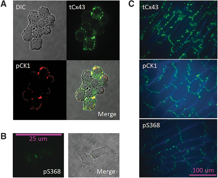
Background: Ischemic preconditioning induces lateralization and dephosphorylation of Connexin 43 (Cx43). However, the Cx43 protein that remains at intercalated disks may be phosphorylated by casein kinase 1 (CK1) and protein kinase C (PKC), and both kinases provide cardioprotection from further ischemic injury. Here we explore the channel characteristics of a Cx43 mutant mimicking preconditioning by CK1 and PKC phosphorylation.

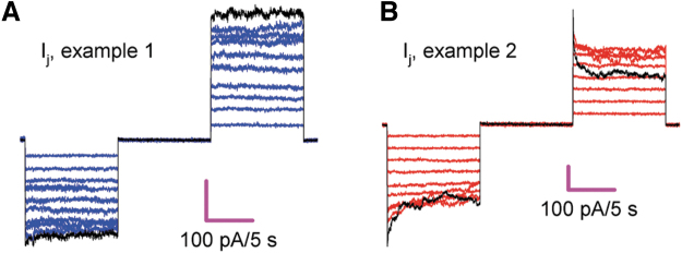
Materials and methods: Whole-cell patch-clamp recordings were performed in cells expressing the mutant Cx43pc (S325,328,330,368D, S365A-Cx43), and the connexin electrical behavior was analyzed at the single channel and macroscopic level.

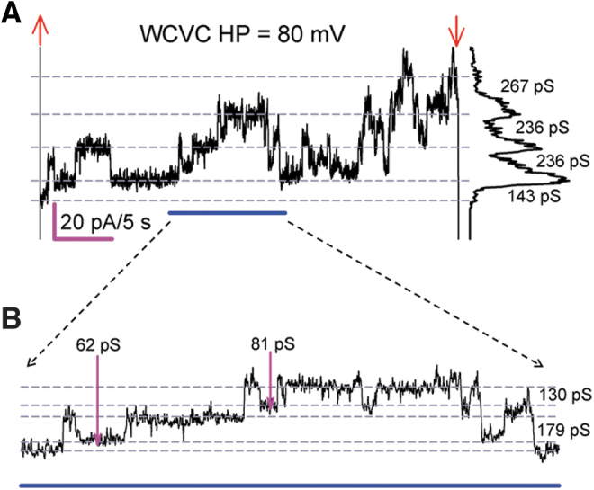
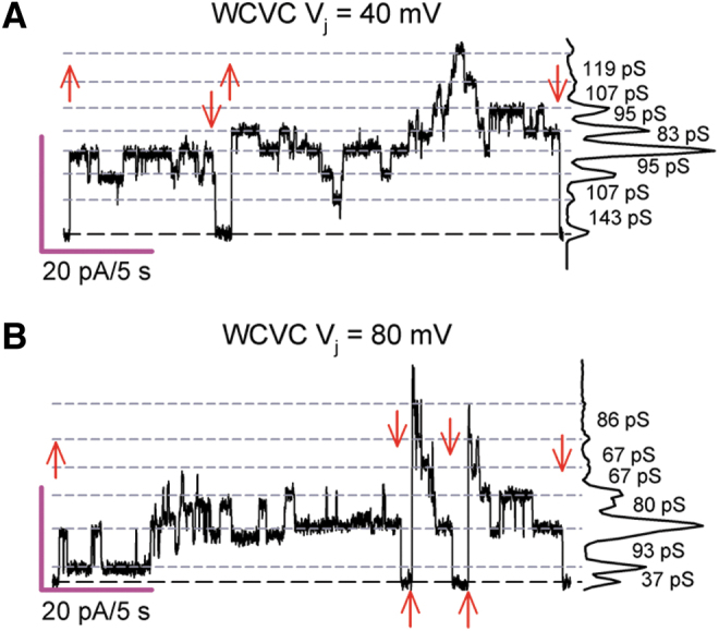
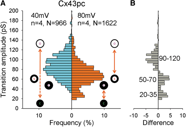
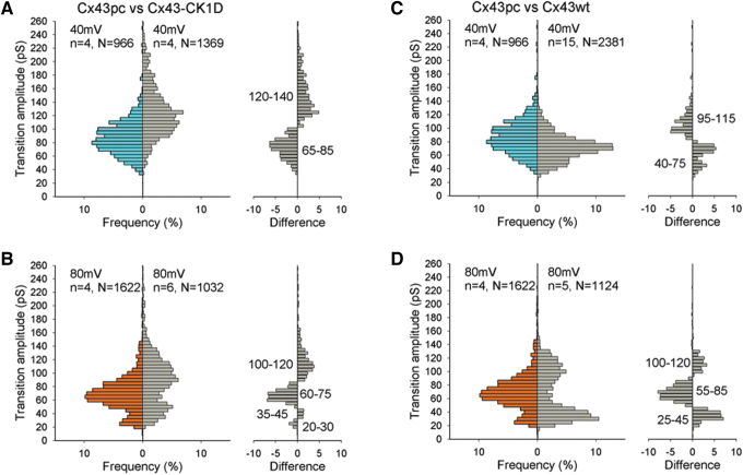
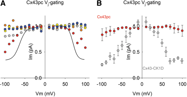
Results: Cx43pc hemichannels opened readily, whereas gap junctions channels displayed amplitudes between the wild-type and CK1 phosphorylated forms, and weaker voltage gating than either counterpart.

Conclusions: Ischemic preconditioning and the ensuing phosphorylation of Cx43 by PKC may render junctional channels insensitive to transjunctional voltages, allowing the preservation of intercellular communication in ischemic conditions.

Keywords: CK1; Cx43; PKC; ischemia; patch clamp; preconditioning.


Conflict of interest statement

No competing financial interests exist.

Figures

FIG. 1.
FIG. 1.
Cx43 is phosphorylated by CK1 and PKC. (A, B) WT Cx43 in Rin GVA cells is abundantly expressed, and immunostaining with phosphospecific antibodies shows that plaque Cx43 (at cell-to-cell contacts) is phosphorylated by CK1 (A) and PKC (B) in cell-to-cell contacts). DIC image; colocalization of tCx43 and Cx43 phosphorylated at CK1 target sites (pCK1) shown in yellow at the merged image in a cell group. For pS368 Cx43, only that specific antibody was used, the merged image is the DIC and the fluorescence. (C) Mouse cardiac tissue after 15 min of global ischemia (see Materials and Methods section) displays lateral relocation of tCx43, and abundant phosphorylation at the CK1 and PKC sites of the protein remaining at the intercalated disks. WGA (blue) stain delineates cell membranes. Cx43 phosphorylated at CK1 and PKC sites appears transverse to the fibers' direction. CK1, casein kinase 1; DIC, differential interference contrast; PKC, protein kinase C; Rin, rat insulinoma; tCx43, total Cx43; WGA, wheat germ agglutinin; WT Cx43, wild-type Cx43.
FIG. 2.
FIG. 2.
Cx43pc HChs open readily. (A) Representative Im sample (37 s) from a single Cx43pc-expressing Rin cell at +80 mV. The cell displayed frequent HCh openings twice the amplitude of GJCh transitions at sustained positive membrane voltage (A). Voltage pulse onset (upward red arrows) and offset (downward red arrows) are shown. The dotted lines indicate the various persistent current levels, and the plot at the right is the all-points histograms with transition conductance (g) values indicated. In (B), an expansion of the underlined (blue) region from (A) reveals the existence of subconductive channel opening (pink arrows). Extracellular Ca2+ = 1 mM.
FIG. 3.
FIG. 3.
GJCh of Cx43pc display smaller transitions at larger Vj values. (A, B) Cx43pc GJCh transitions of variable amplitude suggest multiple conductive states at transjunctional voltage (Vj) of 40 mV (A) and 80 mV (B). Settings as shown in Figure 2. Notice that at Vj = 80 mV, channel transitions denote smaller conductance values than at Vj = 40 mV.
FIG. 4.
FIG. 4.
Cx43pc GJCh transition amplitudes decrease at larger Vj values. (A) Histograms of transition amplitudes from Cx43pc gap junction channels. Channel conductive states (open, substates including a residual) and the nonconductive state (closed) are symbolized to represent the likely transitions between them. (B) Difference plot (transitions at 80 mV minus transitions at 40 mV). Visits to the residual and substates are more frequent, and transitions from the fully open to the fully closed state are less frequent at the higher Vj value.
FIG. 5.
FIG. 5.
GJCh of Cx43pc displays smaller transitions than mutant Cx43–CK1D, and similar transitions to WT Cx43. (A, B) Comparisons of histogram of transitions amplitudes of Cx43pc and Cx43–CK1D show smaller range and smaller transitions for the “preconditioned” mutant, at both Vj = 40 (A) and 80 (B) mV. (C, D) Similar comparisons with WT Cx43 reveal a similar range of amplitudes, but more intermediate states for Cx43pc.
FIG. 6.
FIG. 6.
Cx43pc gap junctions display weak or no Vj-gating sensitivity. (A, B) Two examples of junctional current (Ij) in response to a series of Vj pulses of increasing strength, from 10 to 100 mV, in 10 mV steps. The current at 100 mV is shown in black to accentuate the lack of Vj-gating (A) observed in the group and the only pair that showed Vj-gating (B). To ensure proper voltage control, only pairs with low gj values (mean ± SEM = 4.61 ± 0.63; n = 5) were used for this analysis. SEM, standard error of the mean.
FIG. 7.
FIG. 7.
Cx43pc junctional conductance displays lower Vj-gating than WT Cx43 of Cx43–CK1D. (A, B) Normalized junctional conductance (Gj)/Vj curves from Cx43pc experiments shown in Figure 5 were not amenable to fit with a Boltzmann function (A), in comparison with Cx43wt (black lines; mean fit of published data), or a phosphomutant of only the CK1 sites (B; Cx43–CK1D; see Ref.).

Similar articles

References

    1. Cooper CD, Lampe PD. Casein kinase 1 regulates connexin-43 gap junction assembly. J Biol Chem 2002;277(47):44962–44968. - PubMed
    1. Lampe PD, Cooper CD, King TJ, et al. . Analysis of Connexin43 phosphorylated at S325, S328 and S330 in normoxic and ischemic heart. J Cell Sci.2006;119(Pt 16):3435–3442. - PMC - PubMed
    1. Beardslee MA, Lerner DL, Tadros PN, et al. . Dephosphorylation and intracellular redistribution of ventricular connexin43 during electrical uncoupling induced by ischemia. Circ Res 2000;87(8):656–662. - PubMed
    1. Srisakuldee W, Jeyaraman MM, Nickel BE, et al. . Phosphorylation of connexin-43 at serine 262 promotes a cardiac injury-resistant state. Cardiovasc Res 2009;83(4):672–681. - PubMed
    1. Rong B, Xie F, Sun T, et al. . Nitric oxide, PKC-epsilon, and connexin43 are crucial for ischemic preconditioning-induced chemical gap junction uncoupling. Oncotarget 2016;7(43):69243–69255. - PMC - PubMed

LinkOut - more resources