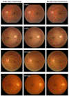
Artificial intelligence, explainability, and the scientific method: A proof-of-concept study on novel retinal biomarker discovery
- PMID: 37746328
- PMCID: PMC10517742
- DOI: 10.1093/pnasnexus/pgad290
Artificial intelligence, explainability, and the scientific method: A proof-of-concept study on novel retinal biomarker discovery
Abstract
We present a structured approach to combine explainability of artificial intelligence (AI) with the scientific method for scientific discovery. We demonstrate the utility of this approach in a proof-of-concept study where we uncover biomarkers from a convolutional neural network (CNN) model trained to classify patient sex in retinal images. This is a trait that is not currently recognized by diagnosticians in retinal images, yet, one successfully classified by CNNs. Our methodology consists of four phases: In Phase 1, CNN development, we train a visual geometry group (VGG) model to recognize patient sex in retinal images. In Phase 2, Inspiration, we review visualizations obtained from post hoc interpretability tools to make observations, and articulate exploratory hypotheses. Here, we listed 14 hypotheses retinal sex differences. In Phase 3, Exploration, we test all exploratory hypotheses on an independent dataset. Out of 14 exploratory hypotheses, nine revealed significant differences. In Phase 4, Verification, we re-tested the nine flagged hypotheses on a new dataset. Five were verified, revealing (i) significantly greater length, (ii) more nodes, and (iii) more branches of retinal vasculature, (iv) greater retinal area covered by the vessels in the superior temporal quadrant, and (v) darker peripapillary region in male eyes. Finally, we trained a group of ophthalmologists () to recognize the novel retinal features for sex classification. While their pretraining performance was not different from chance level or the performance of a nonexpert group (), after training, their performance increased significantly (, ). These findings showcase the potential for retinal biomarker discovery through CNN applications, with the added utility of empowering medical practitioners with new diagnostic capabilities to enhance their clinical toolkit.
Keywords: artificial intelligence; convolutional neural networks; medical image perception; retinal biomarkers; retinal fundus images.
© The Author(s) 2023. Published by Oxford University Press on behalf of National Academy of Sciences.
Figures


References
-
- Suzuki K. 2017. Overview of deep learning in medical imaging. Radiol Phys Technol. 10(3):257–273. - PubMed
LinkOut - more resources
Full Text Sources

