

Radiologic Imaging in Third Nerve Palsy: A Case Series Investigating Etiology, Patterns, and Clinical Implications
- PMID: 37746364
- PMCID: PMC10516257
- DOI: 10.7759/cureus.43986
Radiologic Imaging in Third Nerve Palsy: A Case Series Investigating Etiology, Patterns, and Clinical Implications
Abstract
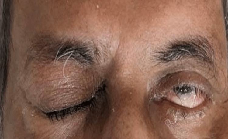
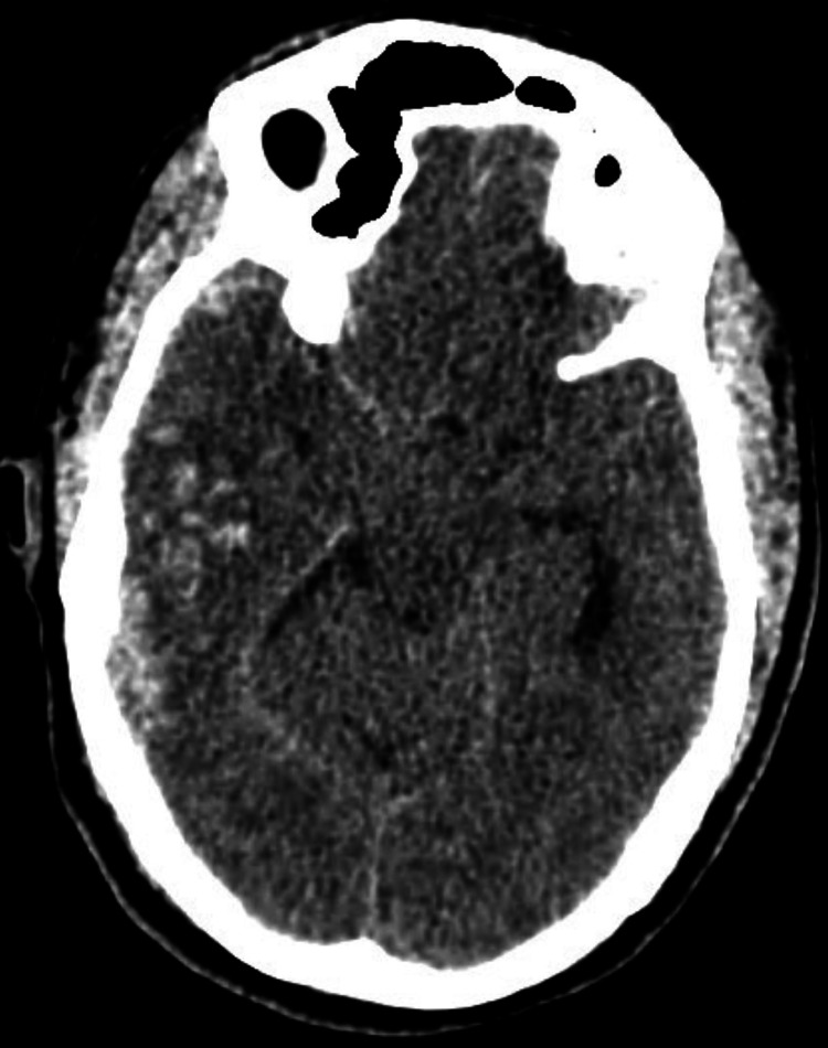
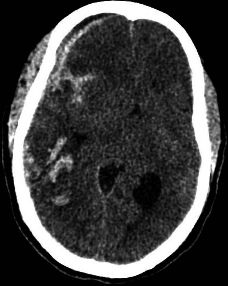
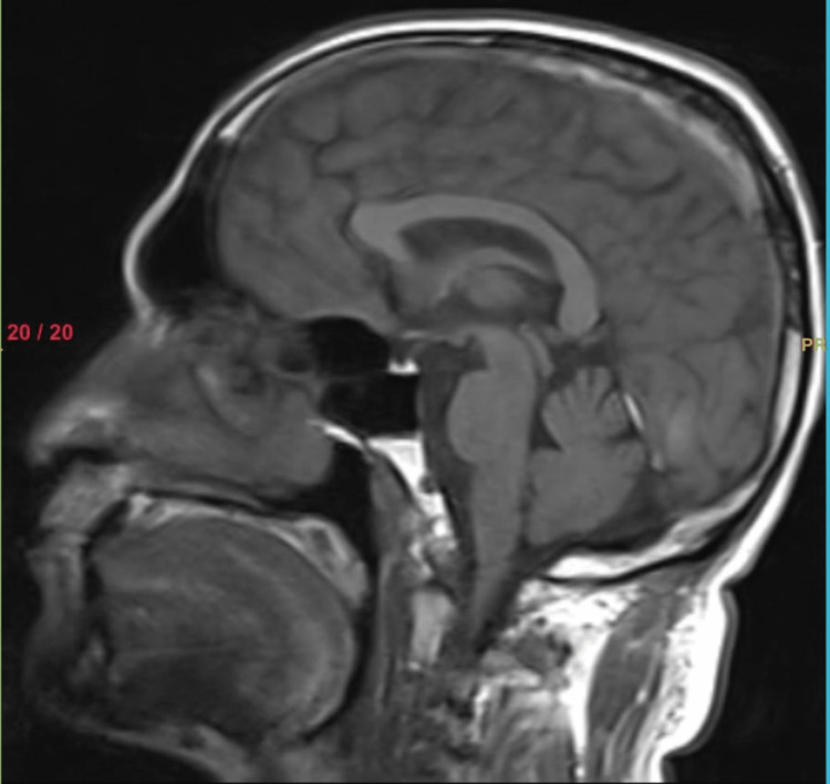
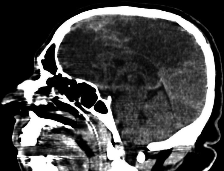
Third nerve palsy (TNP) is a neurologic condition characterized by dysfunction of the oculomotor nerve, leading to various ocular manifestations. Optic nerve evaluation is of utmost important among all cranial nerve palsies affecting the eye. Dysfunction of the third nerve can indicate an underlying neurologic emergency, such as cavernous arteriovenous fistula or giant cell arteritis. Early recognition and prompt treatment are vital in reversing the clinical and visual impairments associated with oculomotor nerve palsy. The typical presentation of isolated TNP involves deviation of the eye in a downward and outward direction, accompanied by ptosis (drooping of the eyelid) and, potentially, pupil involvement. The decision to use vascular imaging is influenced by factors such as age and clinical risk for an aneurysm. If TNP is isolated or partially present with pupil involvement, it suggests compression of the third nerve and necessitates immediate imaging. Given the serious implications of an intracranial aneurysm, physicians often prioritize vascular imaging during the initial evaluation, if available. However, if clinical findings indicate underlying microvascular ischemia, a delay in imaging may be considered. This case series aims to explore the role of radiologic imaging in understanding the etiology, patterns, and clinical implications of TNP.
Keywords: diplopia; imaging; oculomotor nerve; ptosis; third nerve palsy.
Copyright © 2023, Chodvadiya et al.
Conflict of interest statement
The authors have declared that no competing interests exist.
Figures
Similar articles
-
Superior Division Oculomotor Nerve Palsy and Diabetes Mellitus: A Case Report.Cureus. 2025 Apr 20;17(4):e82612. doi: 10.7759/cureus.82612. eCollection 2025 Apr. Cureus. 2025. PMID: 40400818 Free PMC article.
-
Cavernous internal carotid artery aneurysm presenting with ipsilateral oculomotor nerve palsy: A case report.Radiol Case Rep. 2021 Apr 6;16(6):1339-1342. doi: 10.1016/j.radcr.2021.03.008. eCollection 2021 Jun. Radiol Case Rep. 2021. PMID: 33897925 Free PMC article.
-
A Unique Case of Unilateral Oculomotor Nerve Palsy Secondary to Dengue Fever.Cureus. 2023 Feb 21;15(2):e35281. doi: 10.7759/cureus.35281. eCollection 2023 Feb. Cureus. 2023. PMID: 36994298 Free PMC article.
-
Concomitant ectatic posterior communicating artery and tentorial meningioma as a source of oculomotor palsy: case report.Neurosurgery. 2005 Dec;57(6):E1316; discussion E1316. doi: 10.1227/01.neu.0000187448.96386.03. Neurosurgery. 2005. PMID: 16331147 Review.
-
Isolated Ocular Motor Nerve Palsies.Semin Neurol. 2015 Oct;35(5):539-48. doi: 10.1055/s-0035-1563568. Epub 2015 Oct 6. Semin Neurol. 2015. PMID: 26444399 Review.
References
-
- Imaging of oculomotor (third) cranial nerve palsy. Vaphiades MS, Roberson GH. Neurol Clin. 2017;35:101–113. - PubMed
-
- Pathology of the ocular motor nerves III, IV, and VI. Adams ME, Linn J, Yousry I. Neuroimaging Clin N Am. 2008;18:261-82, preceding x-x. - PubMed
-
- Traumatic orbital third nerve palsy. Nagendran ST, Lee V, Perry M. Br J Oral Maxillofac Surg. 2019;57:578–581. - PubMed
-
- Ocular manifestations of head injury and incidence of post-traumatic ocular motor nerve involvement in cases of head injury: a clinical review. Sharma B, Gupta R, Anand R, Ingle R. Int Ophthalmol. 2014;34:893–900. - PubMed
Publication types
LinkOut - more resources
Full Text Sources