

Combining Endovascular Coiling and Open Evacuation for a Delayed-Onset Ruptured Post-traumatic Pseudoaneurysm of the Distal Paracentral Pericallosal Artery Branch
- PMID: 37746416
- PMCID: PMC10511349
- DOI: 10.7759/cureus.43880
Combining Endovascular Coiling and Open Evacuation for a Delayed-Onset Ruptured Post-traumatic Pseudoaneurysm of the Distal Paracentral Pericallosal Artery Branch
Abstract
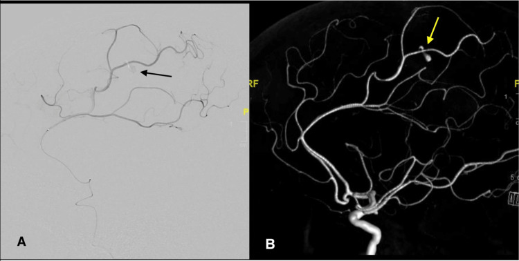
Cerebrovascular pseudoaneurysm development and rupture is a rare, delayed sequelae of trauma. We present a case of a female patient in her sixties who presented after a fall without evidence of vascular injury on imaging. However, after one week, repeat imaging due to an abrupt change in mental status revealed a ruptured pseudoaneurysm, which was treated with a combination of coil embolization and open surgical evacuation of associated intracranial hematoma. This case illustrates the importance of continued surveillance beyond the acute traumatic period to identify late-onset complications in trauma patients requiring emergent treatment.
Keywords: blunt cervical trauma; endovascular coil embolization; frontal craniotomy; intracranial hematoma; traumatic pseudoaneurysm.
Copyright © 2023, Koneru et al.
Conflict of interest statement
The authors have declared that no competing interests exist.
Figures


References
-
- Trauma to the cerebrovascular system. Bula WI, Loes DJ. https://europepmc.org/article/med/7858919. Neuroimaging Clin N Am. 1994;4:753–772. - PubMed
-
- Chronic epidural hematoma caused by traumatic intracranial pseudoaneurysm of the middle meningeal artery: review of the literature with a focus on this unique entity. Umana GE, Cristaudo C, Scalia G, et al. World Neurosurg. 2020;136:198–204. - PubMed
-
- Delayed rupture of traumatic intracranial pseudoaneurysm in a child following gunshot wound to the head. Alvarez JA, Bambakidis N, Takaoka Y. https://europepmc.org/article/med/11951264 J Craniomaxillofac Trauma. 1999;5:39–44. - PubMed
-
- Delayed intracerebral hemorrhage after pseudoaneurysm of middle meningeal artery rupture: case report, literature review, and forensic issues. Montanari E, Polonara G, Montalti R, Vivarelli M, Ricciuti RA, Giorgetti R, Tagliabracci A. World Neurosurg. 2018;117:394–410. - PubMed
Publication types
LinkOut - more resources
Full Text Sources