. 2023 Sep 7:17:1235915.
doi: 10.3389/fncir.2023.1235915. eCollection 2023.

Ultrastructural and immunocytochemical evidence of a colonial nervous system in hydroids

Affiliations

Ultrastructural and immunocytochemical evidence of a colonial nervous system in hydroids

Igor A Kosevich. Front Neural Circuits. .

Abstract

Background: As the sister group to all Bilateria, representatives of the phylum Cnidaria (sea anemones, corals, jellyfishes, and hydroids) possess a recognizable and well-developed nervous system and have attracted considerable attention over the years from neurobiologists and evo-devo researchers. Despite a long history of nervous system investigation in Cnidaria, most studies have been performed on unitary organisms. However, the majority of cnidarians are colonial (modular) organisms with unique and specific features of development and function. Nevertheless, data on the nervous system in colonial cnidarians are scarce. Within hydrozoans (Hydrozoa and Cnidaria), a structurally "simple" nervous system has been described for Hydra and zooids of several colonial species. A more complex organization of the nervous system, closely related to the animals' motile mode of life, has been shown for the medusa stage and a few siphonophores. Direct evidence of a colonial nervous system interconnecting zooids of a hydrozoan colony has been obtained only for two species, while it has been stated that in other studied species, the coenosarc lacks nerves.

Methods: In the present study, the presence of a nervous system in the coenosarc of three species of colonial hydroids - the athecate Clava multicornis, and thecate Dynamena pumila and Obelia longissima - was studied based on immunocytochemical and ultrastructural investigations.

Results: Confocal scanning laser microscopy revealed a loose system composed of delicate, mostly bipolar, neurons visualized using a combination of anti-tyrosinated and anti-acetylated a-tubulin antibodies, as well as anti-RF-amide antibodies. Only ganglion nerve cells were observed. The neurites were found in the growing stolon tips close to the tip apex. Ultrastructural data confirmed the presence of neurons in the coenosarc epidermis of all the studied species. In the coenosarc, the neurons and their processes were found to settle on the mesoglea, and the muscle processes were found to overlay the nerve cells. Some of the neurites were found to run within the mesoglea.

Discussion: Based on the findings, the possible role of the colonial nervous system in sessile hydroids is discussed.

Keywords: Clava multicornis; Cnidaria; Dynamena pumila; Hydrozoa; Obelia longissima; coenosarc; colonial nervous system.


Conflict of interest statement

The author declares that the research was conducted in the absence of any commercial or financial relationships that could be construed as a potential conflict of interest.

Figures

Figure 1
Figure 1
Scheme of the polypoid colony organization in thecate hydroids, and the spatial relationship between the soft tissues and the outer skeleton (perisarc). The colony consists of tubular stolons that grow over the substrate, as well as shoots branching from the stolons, which carry numerous zooids (hydranths). Coenosarc (a tubular tissue composed of two epithelial layers) and hydranths are shown in gray. h, hydranth; hb, hydranth bud (rudiment); ht, hydrotheca; p, perisarc; s, stolon; shgt, shoot growing tip; shin, shoot internode; sgt, stolon growing tip; and sin, stolon internode.
Figure 2
Figure 2
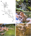
Clava multicornis colony. (A) Scheme of a colony growing on a flat and free substrate. (B) The small colony growing on the brown algae Ascophyllum nodosum. The compact colony occupies the node of the frond. (C) Close view of the interlacing stolons covered with the perisarc, while the hydranths are “naked.” (D, E) Distal parts of stolons growing straight at the periphery of the colony over the free substrate. g, gonads; h, hydranth; hb, hydranth base; s, stolon; and sgt, stolon growing tip. Gray rectangles show the colony parts used for the investigation.
Figure 3
Figure 3
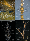
Details of Dynamena pumila (A–C) and Obelia longissima (D, E) colonies. White rectangles indicate the colony elements used for the study. (A) General view of the peripheral part of the D. pumila colony showing the straight-line mode of stolon growth and the regular branching of stolons from the bases of the shoots. (B) Close view of the peripheral part of the colony with young shoots with a monopodial mode of growth. (C) Part of D. pumila shoot illustrating its organization: the two opposite rows of hydrothecae partly fused with the shoot stem; the shoot is subdivided into equal internodes, each composed of a pair of hydrothecae. (D) General view of the characteristic large shoot of O. longissima with regular branching. (E) Close view of a part of a O. longissima shoot depicting the large straight internodes of the stem and the smaller internodes of lateral branches. br_I, the first order branch; br_II, branch of the second order; h, hydranth; ht, hydrotheca; sh, shoot; shgt, shoot growing tip; shin, shoot internode; shst, shoot stem; sgt, stolon growing tip.
Figure 4
Figure 4
Alpha-tubulin-like and anti-RF-amide-like immunoreactivity in the stolon of Clava multicornis. Confocal laser scanning microscopy. Maximum Z-projections (A–E) after immunostaining against alpha-tubulin shown in yellow (A, B, G) or magenta (C, E), and RF-amide shown in white (D, E). Nuclear staining (DAPI) shown in blue (B) and phallacidin staining shown in green (F). (A, B) A straight part of the stolon between the hydranth bases with alpha-tubulin-like immunoreactive cells. (C–E) The same part of the stolon (and the same stack of optical sections) showing overlapping of the alpha-tubulin-like and the anti-RF-amide-like immunoreactivity. (F, G) Part of a stolon with the lateral growing tip; staining with phallacidin (F) helps visualize the outer shape and the borders of the epithelia layers; immunostaining against alpha-tubulin (G) shows neuron-like cells and processes within the new growing tip, as well as numerous nematoblasts. c, cilia; epd, epidermis; gd, gastrodermis; mg, mesoglea; nb, nematoblasts; npr, nerve process; pk, perikaryon; and sgt, stolon growing tip; arrowheads point to the place where the perisarc was disrupted that caused the disturbance of the epidermis integrity. (A–E) Maximum Z-projections of 38 optical sections with 0.3 μm step (overall section thickness-−11.1 μm). (F, G) Maximum Z-projections of 56 optical sections with 0.2 μm step (overall section thickness-−11.0 μm). Scale bar – 50 μm.
Figure 5
Figure 5
Organization of the basal part of the epidermis at the base of the hydranth (A–C) and in the stolon (D) of Clava multicornis, transverse ultrathin sections, TEM. (A) Sheet of epidermal longitudinal muscular processes over the mesoglea at the basal part of the hydranth (the section runs slightly oblique). (B) The enlarged part marked on A showing the neurite bundle. (C) The neurite bundle at the base of the hydranth epidermis separated from the mesoglea by the muscular processes. (D) The neurite bundle in the stolon epidermis in direct contact with the mesoglea and superimposed by muscular processes. epd, epidermis; gd, gastrodermis; mf, muscular microfilaments; mg, mesoglea; mt, microtubules; npr, nerve process; and v, vesicles.
Figure 6
Figure 6
Cross-sections of the Dynamena pumila stolon in different parts. Semi-thin histological sections. (A) A cross-section of the stolon growing tip–soft tissues occupy the entire space within the perisarc tube and have permanent contact with its walls. (B) Cross-section of the stolon between the shoot bases—the coenosarc tube lies on the “bottom” of the perisarc tube and partly spreads on its side walls. epd, epidermis; epl, epidermal layer; gd, gastrodermis; gvc, gastro-vascular cavity; mg, mesoglea; and p, perisarc.
Figure 7
Figure 7
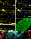
Alpha-tubulin-like immunoreactivity in the stolon of Dynamena pumila. Confocal laser scanning microscopy. Z-projections (A–H) after immunostaining against alpha-tubulin shown in yellow (A–F) or red (G); nuclear staining (DAPI) shown in blue (B, D, F); phallacidin staining shown in cyan (G) or green (H). (A–F) Immunostaining against the alpha-tubulin of different parts of the stolon between the shoot bases: the nuclear staining confirms the visualization of the perikaria. (G) YZ projection (a virtual cross-section of the stolon) of the region in E between dashed lines, including the perikarion of the nerve-like cell [pk in (F)] located at the base of the epidermis over the mesoglea (compare with Figure 6B); yellow dashed circles mark the levels of the mesoglea and the border between the gastrodermis and gastro-vascular cavity. (H) Staining of the stolon growing tip with phallacidin showing the network of the muscle processes. c, cilia of the gastrodermal cells; cmf, circular muscular microfilaments; epd, epidermis; epl, epidermal layer; gd, gastrodermis; gvc, gastro-vascular cavity; lmf, longitudinal muscular microfilaments; mpk, perikaryon of the multipolar labeled cell; nb, nematoblast; npr, neurite-like process; and pk, perikarya of the labeled cells; white asterisks mark the artifacts, non-specific labeling of the traumatized tissue and disrupted perisarc; white arrowheads point to the cilia of the gastrodermal cells. (A, B) Maximum Z-projections of 24 optical sections with 0.15 μm step (overall section thickness −3.45 μm). (C, D) Maximum Z-projections of 36 optical sections with 0.1 μm step (overall section thickness −3.5 μm). (E, F) Maximum Z-projections of 73 optical sections with 0.2 μm step (overall section thickness −14.4 μm). H, maximum Z-projections of 38 optical sections with 0.2 μm step (overall section thickness −7,4 μm). In (E, F), the optical sections pass parallel to the substrate practically through the middle of the stolon perpendicular to its side wall. Scale bars: (A, B, E, F), 50 μm; (C, D, G, H), 20 μm.
Figure 8
Figure 8
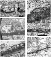
The basal part of the stolon epidermis of Dynamena pumila illustrating some ultrastructural features of the ganglion nerve cells. Longitudinal ultrathin section, TEM. (A) Soma of the neuron located below the muscular processes of epidermal cells and lying over the neurites, close to the mesoglea. (B, C) Enlarged view of the regions marked in A on the left (B) and right (C) sides. (D, E) Bundle of neurites with numerous microtubules and dense-core vesicles. epd, epidermis; gd, gastrodermis; mf, contractile myofibrils; mg, mesoglea; mt, microtubules; n, nucleus; npr, nerve process; and v, vesicles.
Figure 9
Figure 9
Details of the neuron-like cells in the coenosarc of Obelia longissima shoot internode, longitudinal optical (A–C), and ultrathin (D–F) sections (TEM). (A, B) —z-projections after immunostaining against alpha-tubulin [shown in yellow (A, B)] and nuclear staining [shown in blue (B, superimposition with image A)] (confocal laser scanning microscopy); z-projections of 50 optical sections with 0.175 μm step (overall section thickness −8.575 μm). (C) Semi-thin histological section of the part of the internode. (D–F) The ultrastructure of the basal part of the epidermis showing the features of the neurites. epd, epidermis; gd, gastrodermis; mf, contractile myofibrils; mg, mesoglea; mt, microtubules; nb, nematoblast; and npr, nerve process; p, perisarc; pk, perikaryon of the labeled cell; black arrowheads point to the microtubules. Scale bar: a, B−50 μm.
Figure 10
Figure 10
Histological (A) and ultrastructural (B, C) cross-sections of the shoot internode of Obelia longissima. (A) The cross-section of the entire internode; the situation when the specimen was fixed at the time-point when the gastro-vascular cavity was closed due to the periodic peristaltic wave of coenosarc contraction. (B) Neurites located over the mesoglea at the base of the epithelia-muscular epidermal cells. (C) Part of the cross-section of the epidermis base showing the spatial relationship between the neurites and the other cells of the epidermis. emc, epithelia-muscular cell; epd, epidermis; gd, gastrodermis; ic, i-cell; mf, contractile myofibrils; mg, mesoglea; mt, microtubules; nb, nematoblast (with nematocyst); npr, nerve process; and p, perisarc.
Figure 11
Figure 11
Details of the ultrastructure of the neurites visible at the cross-sections of the Obelia longissima shoot internode. Ultrathin sections, TEM. (A–C) A separate neurite (A) and a small bunch of neurites (B, C) showing the characteristic features of the nerve processes: the microtubules and the dense-core vesicles. (D) A small bunch of neurites submerged in mesoglea. (E, F) Flattened bundles of neurites between the mesoglea and the base of the epidermal cells. White asterisk marks the probable neurite submerged in mesoglea. emc, epithelia-muscular cell; epd, epidermis; gd, gastrodermis; mf, contractile myofibrils; mg, mesoglea; mt, microtubules; npr, nerve process; and v, vesicles.

Similar articles

References

    1. Aizenshtadt T. B., Polteva D. G. (1981). Origin of germ cells and early stages of oogenesis in marine hydroid polyp obelia. Ontogenez 12, 243–250.
    1. Anderson P. A. V. (2004). Cnidarian neurobiology: what does the future hold? Hydrobiologia 530, 107–116. 10.1007/s10750-004-2660-x - DOI
    1. Antsulevich A. E. (2015). Hydrozoa (Hydroids and Hydromedusae) of Russian seas. St. Petersburg: St. Petersburg University Press.
    1. Beloussov L. V. (1973). Growth and morphogenesis of some marine hydrozoa according to histological data and time-lapse studies. Publ. Seto Mar. Biol. Lab. 20, 315–366.
    1. Beloussov L. V. (1991). Basic morphogenetic processes in hydrozoa and their evolutionary implications: an exercise in rational taxonomy. Hydrobiologia, 216/217, 61–67. 10.1007/BF00026444 - DOI

Publication types