

Case Reports
doi: 10.1016/j.hrcr.2023.07.002.
eCollection 2023 Sep.
Successful catheter ablation for verapamil-sensitive idiopathic left ventricular tachycardia guided by dual post-QRS wave P1 potentials after catheter-induced mechanical block
Affiliations
- PMID: 37746557
- PMCID: PMC10511895
- DOI: 10.1016/j.hrcr.2023.07.002
Item in Clipboard
Case Reports
Successful catheter ablation for verapamil-sensitive idiopathic left ventricular tachycardia guided by dual post-QRS wave P1 potentials after catheter-induced mechanical block
HeartRhythm Case Rep.
.
No abstract available
Keywords: Bump phenomenon; Catheter-induced mechanical block; Late P1 potential; Playback ablation; Verapamil-sensitive left fascicular ventricular tachycardia.
Figures
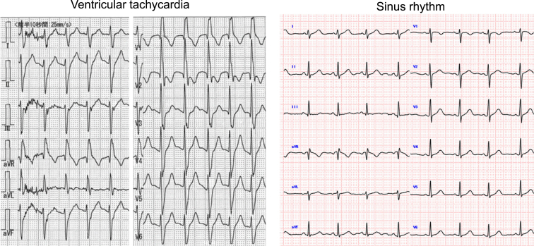
Twelve-lead electrocardiogram (left, ventricular tachycardia; right, sinus rhythm). HIS = bundle of His; HRA = high right atrium; LAO, left anterior oblique; RV = right ventricle.
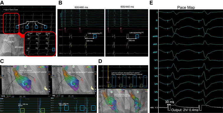
A: Intracardiac electrogram and fluoroscopic image at the moment of the “bump” phenomenon. B: Atrial programmed pacing that demonstrates decremental conduction in the late-appearing P1 potential. C: Two types of delayed P1 potentials, recorded by the DECANAV (DECA) catheter in the CARTO3 system (Biosense Webster, Diamond Bar, CA). The left panel displays the site recorded for the early-to-mid-appearing P1 potential, marked with a light blue tag, while the right panel displays the site recorded for the late-appearing P1 potential, marked with a green tag. D: The upper panel displays the local potentials of the ablation catheter at the beginning of radiofrequency ablation. The presumed bump site is identified by the blue tag and the ablation sites are identified by red or pink tags in the lower panel. E: Best pace map at the ablation site.
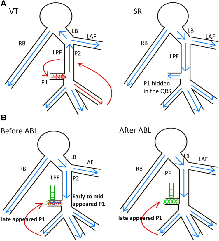
A: Illustration showing the appearance of the P1 potential in typical idiopathic left ventricular tachycardia (left, ventricular tachycardia; right, sinus rhythm). B: Illustration showing the mechanisms that cause the 2 types of delayed P1 potentials (left, before ablation; right, after ablation). ABL = ablation; LAF = left anterior fascicle; LB = left bundle; LPF = left posterior fascicle; RB = right bundle; SR = sinus rhythm; VT = ventricular tachycardia.
Similar articles
-
A case with occurrence of antidromic tachycardia after ablation of idiopathic left fascicular tachycardia: mechanism of left upper septal ventricular tachycardia.J Cardiovasc Electrophysiol. 2013 Jul;24(7):825-7. doi: 10.1111/jce.12072. Epub 2013 Jan 25. J Cardiovasc Electrophysiol. 2013. PMID: 23350939
-
Left posterior fascicular block: a new endpoint of ablation for verapamil-sensitive idiopathic ventricular tachycardia.Chin Med J (Engl). 2006 Mar 5;119(5):367-72. Chin Med J (Engl). 2006. PMID: 16542578
-
Verapamil-sensitive left anterior fascicular ventricular tachycardia: results of radiofrequency ablation in six patients.J Cardiovasc Electrophysiol. 1998 Dec;9(12):1269-78. doi: 10.1111/j.1540-8167.1998.tb00102.x. J Cardiovasc Electrophysiol. 1998. PMID: 9869526
-
Idiopathic left ventricular tachycardia: assessment and treatment.Card Electrophysiol Rev. 2002 Dec;6(4):448-57. doi: 10.1023/a:1021100828459. Card Electrophysiol Rev. 2002. PMID: 12438827 Review.
-
[Fascicular ventricular tachycardia].Ital Heart J Suppl. 2001 Nov;2(11):1181-6. Ital Heart J Suppl. 2001. PMID: 11775409 Review. Italian.
References
-
- Zipes D.P., Foster P.R., Troup P.J., Pedersen D.H. Atrial induction of ventricular tachycardia: reentry versus triggered automaticity. Am J Cardiol. 1979;44:1–8. - PubMed
-
- Klein G.J., Millman P.J., Yee R. Recurrent ventricular tachycardia responsive to verapamil. Pacing Clin Electrophysiol. 1984;7:938–948. - PubMed
-
- Okumura K., Matsuyama K., Miyagi H., Tsuchiya T., Yasue H. Entrainment of idiopathic ventricular tachycardia of left ventricular origin with evidence for reentry with an area of slow conduction and effect of verapamil. Am J Cardiol. 1988;62:727–732. - PubMed
-
- Nogami A., Naito S., Tada H., et al. Demonstration of diastolic and presystolic Purkinje potentials as critical potentials in a macroreentry circuit of verapamil-sensitive idiopathic left ventricular tachycardia. J Am Coll Cardiol. 2000;36:811–823. - PubMed
-
- Liu Q., Shehata M., Jiang R., et al. Macroreentrant loop in ventricular tachycardia from the left posterior fascicle: new implications for mapping and ablation. Circ Arrhythm Electrophysiol. 2016;9 - PubMed
Publication types
LinkOut - more resources
Full Text Sources
