. 2023;43(10):515-530.
doi: 10.1080/10985549.2023.2253130. Epub 2023 Oct 11.

Module 4-Deficient CCN2/Connective Tissue Growth Factor Attenuates the Progression of Renal Fibrosis via Suppression of Focal Adhesion Kinase Phosphorylation in Tubular Epithelial Cells

Affiliations

Module 4-Deficient CCN2/Connective Tissue Growth Factor Attenuates the Progression of Renal Fibrosis via Suppression of Focal Adhesion Kinase Phosphorylation in Tubular Epithelial Cells

Hiroaki Amano et al. Mol Cell Biol. 2023.

Abstract

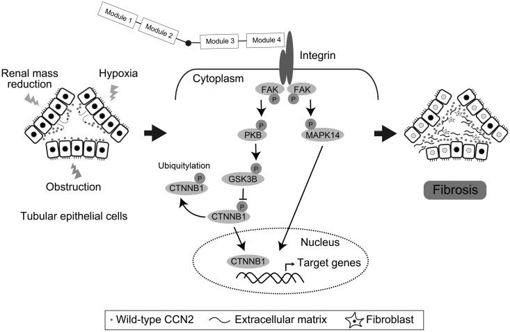
CCN2/connective tissue growth factor (CTGF) potentially serves as a therapeutic target for chronic kidney disease. Here we investigated CCN2 module-4, encoded by Ccn2 exon 5, through the generation of Ccn2 exon 5 knockout mice (Ex5-/- mice). To investigate renal fibrosis pathogenesis, Ex5-/- mice were employed to model unilateral ureteral obstruction (UUO), unilateral ischemic-reperfusion injury (UIRI), and 5/6 nephrectomy. Interstitial fibrosis was significantly attenuated in the Ex5-/- mice in the three models. Furthermore, phosphorylated focal adhesion kinase (FAK) levels in tubular epithelial cells were significantly lower in the kidneys of the UUO- and UIRI-Ex5-/- mice than those of the Ex5+/+ mice. Moreover, CCN2 module 4-mediated renal tubule FAK and promoted fibrosis. These findings indicate that CCN2 module-4-FAK pathway components will serve as therapeutic targets for effectively attenuating renal fibrosis.

Keywords: CCN2/CTGF; CKD; Chronic kidney disease; FAK; Focal adhesion kinase; centralized communication network 2; fibrosis.


Conflict of interest statement

No potential conflict of interest was reported by the author(s).

Figures

FIG 1
FIG 1
Exon 5-deleted Ccn2 knock-in mice develop normally. (A) Genomic maps of wild-type and exon 5-deleted Ccn2. (B) Genotyping PCR of wild-type and exon 5-deleted Ccn2. (C) Western blot analysis of CCN2 expression in lysates of embryonic fibroblasts prepared from Ex5+/+, Ex5+/−, and Ex5−/− mice. Western blots were probed with anti-CCN2 antibody. (D) Protocols for the generation of chronic kidney disease (CKD) models. Ex, exon; Ab, antibody; GAPDH, glyceraldehyde-3-phosphate dehydrogenase; UUO, unilateral ureteral obstruction; UIRI, unilateral ischemia-reperfusion injury; 5/6Nx, 5/6 nephrectomy; mCCN2Δ4, mutant CCN2 lacking module-4.
FIG 2
FIG 2
Interstitial fibrosis is attenuated in CKD models employing exon 5-deleted Ccn2 knock-in mice (Ex5−/− mice). (A–F, H–K, N, O) Masson’s trichrome staining of kidney tissues of UUO, UIRI, and 5/6 Nx model mice. Scale bar = 200 µm. (G, L, P) Extracellular matrix (ECM) deposition (blue area) is shown. Data were obtained from two independent fields of five kidneys and presented as the mean ± standard error of the mean (n = 5 per group). *P < 0.05: the difference is significant compared to Ex5+/+ for the same days. (M) Semiquantitative analysis of the degree of tubular epithelial cell damage. Ex, exon; UUO, unilateral ureteral obstruction; UIRI, unilateral ischemia-reperfusion injury; 5/6Nx, 5/6 nephrectomy.
FIG 3
FIG 3
Expression of mRNAs encoding profibrotic genes decrease in day 7-UUO kidneys of Ex5−/− mice (A–D) qRT-PCR analysis of the expression levels of profibrotic genes in the UUO kidneys of Ex5−/− mice. The mRNA levels of collagen type 1 α1 (Col1a1) and plasminogen activator inhibitor-1 (Pai-1) were significantly lower in day 7-UUO-Ex5−/− kidneys than those of Ex5+/+ mice. (E and F) The levels of wild-type Ccn2 mRNAs amplified using Ex1-2 and Ex4-5 primer sets increased until the date of final analysis. The levels of mRNAs from exon 5-deleted Ccn2 amplified using only the Ex1-2 primer set were higher in Ex5−/− mice than in Ex5+/+ mice. Gapdh mRNA was used as a reference. Values represent the mean ± standard error of the mean (n = 8, per group). *P < 0.05: the difference is significant compared to Ex5+/+ for the same days. qRT-PCR, real-time semiquantitative reverse transcription-polymerase chain reaction; Tgfb1, transforming growth factor-β1; UUO, unilateral ureteral obstruction; Ex, exon; Fn-EIIIA, fibronectin1 EIIIA isoform; Gapdh, glyceraldehyde-3-phosphate dehydrogenase.
FIG 4
FIG 4
Expression of phosphorylated-FAK in day 7-UUO kidneys of Ex5−/− mice. (A and B) Western blot analysis of the expression of phosphorylated-FAK (Tyr397), FAK, and GAPDH in extracts prepared from whole kidneys of day 7-UUO-Ex5−/− and Ex5+/+ mice. GAPDH served as a loading control. Phosphorylation of FAK (Tyr397) was significantly reduced in UUO-Ex5−/− kidneys compared with those of Ex5+/+ kidneys. (C to F) Western blots showing the levels of p-SMAD2 (Ser465/467)/SMAD3 (Ser423/425) and p-LRP6 (Ser1490). Data are presented as mean ± standard error of the mean (n = 5, per group). A significant difference is indicated by *P < 0.05: the difference is significant compared to Ex5+/+. FAK, focal adhesion kinase; Ex, exon; GAPDH, glyceraldehyde-3-phosphate dehydrogenase; UUO, unilateral ureteral obstruction.
FIG 5
FIG 5
Signal transducers downstream of CCN2 M4-FAK pathway in day 7-UUO kidneys. (A) Western blot analysis of the expression of phosphorylated signal transducers in extracts prepared from day 7-UUO kidneys of Ex5−/− and Ex5+/+ mice. GAPDH served as a loading control. (B) Levels of signal transducers downstream of CCN2 M4. The levels p-FAK (Tyr397), p-PI3K p85 (Tyr458)/p55 (Tyr199), and p-PKB (Ser473) were significantly lower in day 7-UUO kidneys of Ex5−/− mice compared with those of Ex5+/+ mice. (C) Western blot analysis of the expression of p-MAPK1/3 (Thr202/Tyr204), p-MAPK8 (Thr183/Tyr185), p-MAPK14 (Thr180/Tyr182), and GAPDH in extracts prepared from kidneys describe above. (D) The levels of p-MAPK1/3 (Thr202/Tyr204) and MAPK1/3 and p-MAPK14 (Thr180/Tyr182) and MAPK14 were significantly lower in day 7-UUO kidneys of Ex5−/− mice compared with those of Ex5+/+ mice. Data are presented as mean ± standard error of the mean (n = 5, per group). Significant differences between Ex5−/− and Ex5+/+ mice are indicated by *P < 0.05. FAK, focal adhesion kinase; M4, module 4; FAK, focal adhesion kinase; UUO, unilateral ureteral obstruction; Ex, exon; GAPDH, glyceraldehyde-3-phosphate dehydrogenase, PI3K: phosphoinositide-3-kinase; PKB: protein kinase B; GSK3B: glycogen synthase kinase 3 beta; MAPK1/3, mitogen-activated protein kinase 1/3, also known as ERK1/2; MAPK8, mitogen-activated protein kinase 8, also known as JNK; MAPK14, mitogen-activated protein kinase 14, also known as p38.
FIG 6
FIG 6
Phosphorylation of FAK and increased levels of CTNNB1 in tubular epithelial cells in mouse models of CKD. (A–J) Immunofluorescence analysis of p-FAK (Tyr397) in of Ex5−/− and Ex5+/+ CKD-model mice. Cells expressing p-FAK and nuclei are stained red and green, respectively. In the control kidneys, p-FAK-positive cells were not detected. In Ex5+/+ mice with CKD, p-FAK (Tyr397)-positive cells increased over time, although few p-FAK (Tyr397)-positive cells were detected in Ex5−/− mice. (K–T) CTNNB1-positive cells were red and nuclei were green. In control kidneys, CTNNB1 localized to adherens junctions in the tubules and accumulated in the cytoplasm of tubular epithelial cells in day 7-UUO and day 14-UIRI kidneys of Ex5+/+ mice. In contrast, few CTNNB1-positive cells were detected in Ex5−/− mice with CKD. (U and V) Quantification of p-FAK (Tyr397)-positive cells in mice with CKD. The p-FAK (Tyr397)-positive cells were detected in the day 7-UUO and day 14-UIRI kidneys of Ex5+/+ mice compared with those of Ex5−/− mice. (W and X) CTNNB1-positive cells were detected in the day 7-UUO and day 14-UIRI Ex5+/+ mice compared with the Ex5−/− mice. Data were obtained from two independent fields of 5 kidneys and are presented as the mean ± standard error of the mean. *P < 0.05: significant difference compared to Ex5+/+. Scale bar = 50 µm. FAK, focal adhesion kinase; CKD, chronic kidney disease; Ex, exon; UUO, unilateral ureteral obstruction; UIRI, unilateral ischemia-reperfusion injury; p-, phosphorylated; HPF, high power field.
FIG 7
FIG 7
Mechanism of the profibrogenic action of CCN2 in the kidneys. Obstruction, hypoxia, and renal mass reduction lead to stimulation of CCN2 secretion from tubular epithelium cells. In the CKD models, CCN2 drives renal fibrogenesis via CCN2 module-4, which stimulates phosphorylation of FAK (Tyr397) in tubular epithelium cells. The synthesis of CCN2 influences the CCN2 module-4-Integrin-FAK pathway by functioning in an autocrine or paracrine manner to promote extracellular matrix deposition in the renal interstitium. CKD, chronic kidney disease; FAK, focal adhesion kinase.

References

    1. Djudjaj S, Boor P.. Cellular and molecular mechanisms of kidney fibrosis. Mol Aspects Med. 2019;65:16–36. doi:10.1016/j.mam.2018.06.002. - DOI - PubMed
    1. Wright JR, Duggal A, Thomas R, Reeve R, Roberts IS, Kalra PA.. Clinicopathological correlation in biopsy-proven atherosclerotic nephropathy: implications for renal functional outcome in atherosclerotic renovascular disease. Nephrol Dial Transplant. 2001;16:765–770. doi:10.1093/ndt/16.4.765. - DOI - PubMed
    1. Ruiz-Ortega M, Rayego-Mateos S, Lamas S, Ortiz A, Rodrigues-Diez RR.. Targeting the progression of chronic kidney disease. Nat Rev Nephrol. 2020;16:269–288. doi:10.1038/s41581-019-0248-y. - DOI - PubMed
    1. Nagai Y, Matoba K, Kawanami D, Takeda Y, Akamine T, Ishizawa S, Kanazawa Y, Yokota T, Utsunomiya K, Nishimura R.. ROCK2 regulates TGF-beta-induced expression of CTGF and profibrotic genes via NF-kappaB and cytoskeleton dynamics in mesangial cells. Am J Physiol Renal Physiol. 2019;317:F839–F851. doi:10.1152/ajprenal.00596.2018. - DOI - PubMed
    1. Yokoi H, Mukoyama M, Sugawara A, Mori K, Nagae T, Makino H, Suganami T, Yahata K, Fujinaga Y, Tanaka I, et al. . Role of connective tissue growth factor in fibronectin expression and tubulointerstitial fibrosis. Am J Physiol Renal Physiol. 2002;282:F933–42. doi:10.1152/ajprenal.00122.2001. - DOI - PubMed

LinkOut - more resources