. 2023 Oct 31;14(5):e0120623.
doi: 10.1128/mbio.01206-23. Epub 2023 Sep 25.

Broad and potent neutralizing antibodies are elicited in vaccinated individuals following Delta/BA.1 breakthrough infection

Affiliations

Broad and potent neutralizing antibodies are elicited in vaccinated individuals following Delta/BA.1 breakthrough infection

Jeffrey Seow et al. mBio. .

Abstract

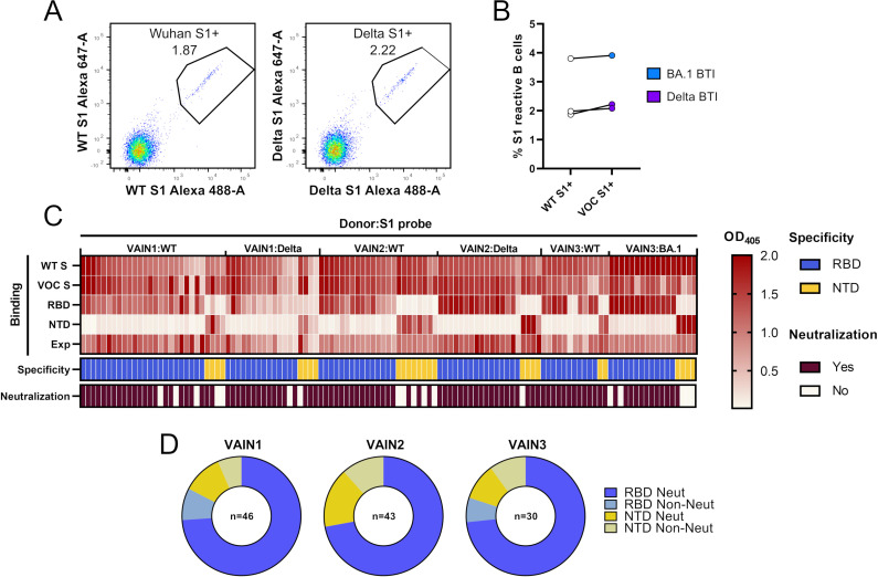
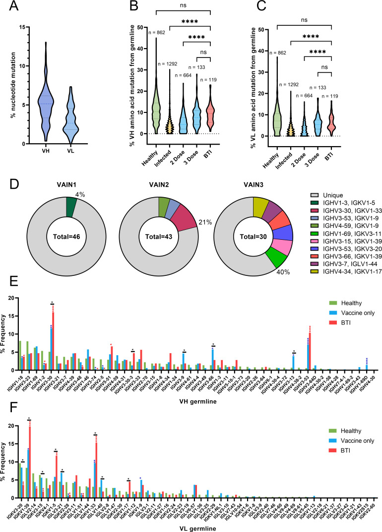
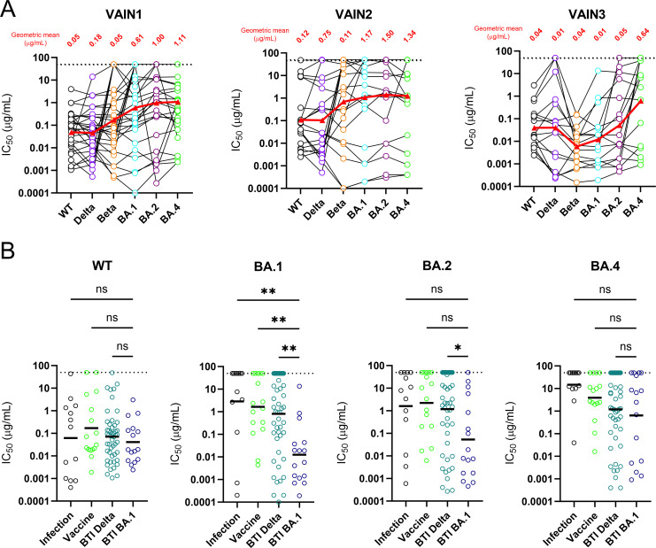
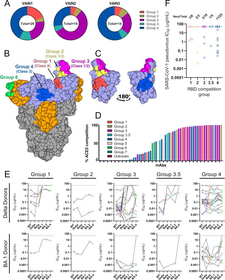
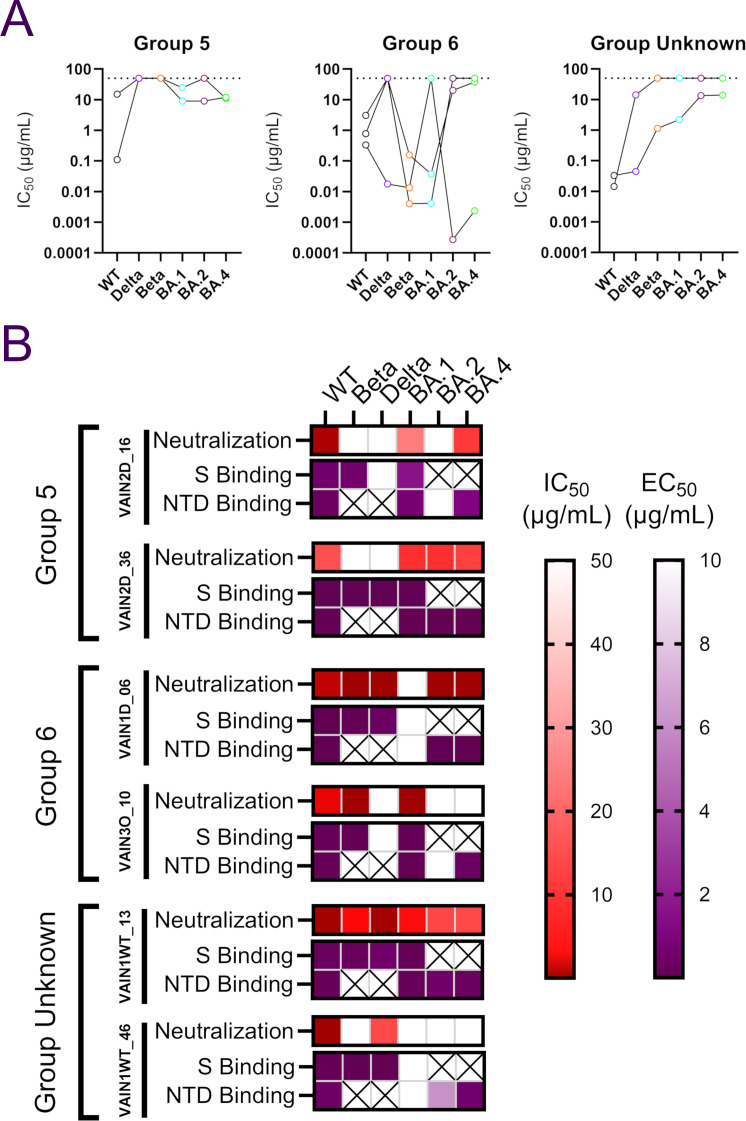
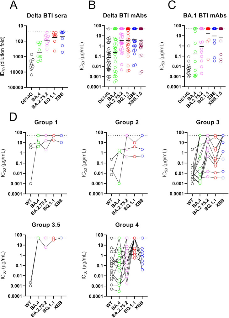
With the emergence of SARS-CoV-2 viral variants, there has been an increase in infections in vaccinated individuals. Here, we isolated monoclonal antibodies (mAbs) from individuals experiencing a breakthrough infection (Delta or BA.1) to determine how exposure to a heterologous Spike broadens the neutralizing antibody response at the monoclonal level. All mAbs isolated had reactivity to the Spike of the vaccine and infection variant. While many mAbs showed reduced neutralization of current circulating variants, we identified mAbs with broad and potent neutralization of BA.2.75.2, XBB, XBB.1.5, and BQ.1.1 indicating the presence of conserved epitopes on Spike. These results indicate that variant-based vaccine boosters have the potential to broaden the vaccine response.

Keywords: SARS-CoV-2; immunization; infectious disease; monoclonal antibodies; neutralizing antibodies.


Conflict of interest statement

The authors declare no conflict of interest.

Figures

Fig 1
Fig 1
Isolation of mAbs using antigen-specific B cell sorting. (A) CD14/CD3/CD8/CD19+/IgM/IgD/IgG+ and S1+ B cells were sorted into 96-well plates. Example: fluorescent-activated cell sorting showing percentage of CD19+IgG+ B cells binding to S1 of Wuhan-1 or S1 of Delta VOC. Full sorting gating strategy is shown in Fig. S1. (B) Percentage of CD19+IgG+ S1 Wuhan and S1 VOC reactive B cells for each donor (Delta for VAIN1 and VAIN2, BA.1 for VAIN3). Data points from the same individuals are linked. Blue: BA.1/Omicron infected and purple: Delta infected. (C) Heatmap showing IgG expression level and binding to SARS-CoV-2 Spike [WT and VOC (Delta for VAIN1 and VAIN2, BA.1 for VAIN3)], and to Spike domains RBD and NTD. The figure reports optical density (OD) values from a single experiment (range 0–2.0) for undiluted supernatant from small-scale transfection of 119 cloned mAbs. Antigen binding was considered positive when OD at 405 nM was >0.2 after subtraction of the background. SARS-CoV-2 Spike domain specificity (RBD or NTD) for each antibody is indicated. Neutralization activity was measured against wild-type (Wuhan) pseudotyped virus using concentrated supernatant and neutralization status is indicated. Antigen probe used to select specific B cells is indicated (i.e., WT S1, Delta S1, or BA.1 S1). (D) Distribution of mAbs targeting RBD and NTD for each donor, as well as their neutralization capability. mAbs are classified as shown in the key (related to Fig. S1 and S2; Table S1).
Fig 2
Fig 2
BTI mAbs show higher somatic hypermutation than mAbs isolated following two vaccine doses. (A) Truncated violin plot showing the percentage of nucleotide mutation compared with germline for the VH and VL genes of Spike-reactive mAbs isolated from VAIN1, VAIN2, and VAIN3. Truncated violin plot comparing the percentage of amino acid mutation compared with germline for (B) VH and (C) VL between Spike-reactive mAbs isolated following infection, two doses of vaccine, three doses of vaccine, or following BTI and IgG B cell receptors (BCRs) from SARS-CoV-2-naive individuals (36). D’Agostino and Pearson tests were performed to determine normality. Based on the result, a Kruskal-Wallis test with Dunn’s multiple comparison post hoc test was performed. *P < 0.0332, **P < 0.0021, ***P < 0.0002, and ****P < 0.0001. (D) Pie chart showing the distribution of heavy chain sequences for donors VAIN1, VAIN2, and VAIN3. The number inside the circle represents the number of heavy chains analyzed. The Pie slice size is proportional to the number of clonally related sequences and is color coded based on clonal expansions described in Table S2. The percentage (%) on the outside of the Pie slice represents the overall % of sequences related to a clonal expansion. Graph showing the relative abundance of (E) V H and (F) V L gene usage for Spike-reactive mAbs isolated following infection (n = 1,292), vaccination (n = 817, including two and three vaccine doses) or following BTI (n = 106), and IgG BCRs from SARS-CoV-2-naive individuals (n = 1,292) (36). Statistical significance was determined by binomial test. *P ≤ 0.05, **P ≤ 0.01, ***P ≤ 0.001, and ****P ≤ 0.0001. Blue stars represent vaccine vs healthy, red stars represent BTI vs healthy, and black stars represent BTI vs vaccine (related to Fig. S3; Table S2).
Fig 3
Fig 3
Neutralization breadth and potency against Omicron sub-lineages. (A) Neutralization breadth and potency of BTI mAbs against Wuhan-1, Delta, Beta, BA.1, BA.2, and BA.4 from VAIN1, VAIN2, and VAIN3. Data for each mAb are linked. Red triangle and linking line show the geometric mean IC50 against each variant. Dotted line represents the highest concentration of antibody tested. (B) Comparison of neutralization breadth and potency of BTI mAbs with mAbs isolated from convalescent donors (infection) (5) and an AZD1222 vaccinated donor (6) against Omicron sub-lineages (BA.1, BA.2, and BA.4). Horizontal line represents the geometric mean IC50 against each mAb origin. D’Agostino and Pearson tests were performed to determine normality. Based on the result, a Kruskal-Wallis test with Dunn’s multiple comparison post hoc test was performed. *P < 0.0332, **P < 0.0021, ***P < 0.0002, and ****P < 0.0001 (related to Table S3).
Fig 4
Fig 4
RBD mAb characterization. (A) Pie chart showing distribution of RBD-specific mAbs between competition groups for VAIN1, VAIN2, and VAIN3. (B) Surface representation of SARS-CoV-2 WT spike (pdb:6XM0) showing epitopes of previously characterized competition groups as colored surfaces (6). RBD competition groups are also shown as RBD classes as defined by Barnes et al. (40). RBD and NTD are indicated by light blue and orange, respectively. (C) Surface representation of RBD domain in the up conformation showing location and proximity of Group 1 (red), Group 2 (yellow), Group 3 (magenta), and Group 4 (blue). Structures were generated in Pymol. (D) Ability of RBD-specific neutralizing antibodies to inhibit the interaction between cell surface ACE2 and soluble SARS-CoV-2 Spike. mAbs (at 600 nM) were pre-incubated with fluorescently labeled Spike before addition to HeLa-ACE2 cells. The percentage reduction in mean fluorescence intensity is reported. Experiments were performed in duplicate. Bars are color coded based on their competition group. (E) Neutralization breadth and potency of RBD-specific mAbs within the different RBD competition groups. mAbs are separated by the infecting VOC. Data for each mAb are linked. Dotted line represents the highest concentration of antibody tested. (F) Neutralization potency of RBD-specific mAbs against SARS-CoV-1. Data are presented by RBD competition group (related to Fig. S4 through S6).
Fig 5
Fig 5
NTD mAb characterization. (A) Neutralization breadth and potency of NTD-specific mAbs within the different NTD competition groups. Data for each mAb are linked. Dotted line represents the highest concentration of antibody tested. (B) Comparison between neutralization activity (IC50) and binding to Spike or NTD (EC50) by ELISA for NTD-specific mAbs. IC50 and EC50 (half-maximal effective concentrations of binding) values are shown as a heat map for each NTD-specific mAb with the level of binding shown in the key. A cross indicates that the Spike or NTD antigen for that variant was not available to test (related to Fig. S5).
Fig 6
Fig 6
mAb neutralization against more recent VOCs including BA.2.75.2, XBB/XBB.1.5, and BQ.1.1. Neutralization by (A) plasma from individuals vaccinated with two doses of BNT162b2 and were subsequently Delta infected (ID50), (B) mAbs from Delta BTI donors (IC50), and (C) mAbs from BA.1 BTI donor (IC50). Sera were collected 15–35 days post-infection. Additional plasma/sera samples from double vaccinated and BA.1-infected individuals were not available. Horizontal line represents geometric mean IC50 (for mAbs) or geometric mean titers (sera). (D) Neutralization breadth and potency broken down by RBD competition group. Data for each mAb are linked. Dotted line represents the highest concentration of mAb or lowest dilution of sera tested (related to Fig. S7).

References

    1. Goldblatt D, Alter G, Crotty S, Plotkin SA. 2022. Correlates of protection against SARS-CoV-2 infection and COVID-19 disease. Immunol Rev 310:6–26. doi:10.1111/imr.13091 - DOI - PMC - PubMed
    1. Addetia A, Crawford KHD, Dingens A, Zhu H, Roychoudhury P, Huang M-L, Jerome KR, Bloom JD, Greninger AL. 2020. Neutralizing antibodies correlate with protection from SARS-CoV-2 in humans during a fishery vessel outbreak with a high attack rate. J Clin Microbiol 58:e02107-20. doi:10.1128/JCM.02107-20 - DOI - PMC - PubMed
    1. Khoury DS, Cromer D, Reynaldi A, Schlub TE, Wheatley AK, Juno JA, Subbarao K, Kent SJ, Triccas JA, Davenport MP. 2021. Neutralizing antibody levels are highly predictive of immune protection from symptomatic SARS-CoV-2 infection. Nat Med 27:1205–1211. doi:10.1038/s41591-021-01377-8 - DOI - PubMed
    1. Lustig Y, Sapir E, Regev-Yochay G, Cohen C, Fluss R, Olmer L, Indenbaum V, Mandelboim M, Doolman R, Amit S, Mendelson E, Ziv A, Huppert A, Rubin C, Freedman L, Kreiss Y. 2021. BNT162b2 COVID-19 vaccine and correlates of humoral immune responses and dynamics: a prospective, single-centre, longitudinal cohort study in health-care workers. Lancet Respir Med 9:999–1009. doi:10.1016/S2213-2600(21)00220-4 - DOI - PMC - PubMed
    1. Graham C, Seow J, Huettner I, Khan H, Kouphou N, Acors S, Winstone H, Pickering S, Galao RP, Dupont L, Lista MJ, Jimenez-Guardeño JM, Laing AG, Wu Y, Joseph M, Muir L, van Gils MJ, Ng WM, Duyvesteyn HME, Zhao Y, Bowden TA, Shankar-Hari M, Rosa A, Cherepanov P, McCoy LE, Hayday AC, Neil SJD, Malim MH, Doores KJ. 2021. Neutralization potency of monoclonal antibodies recognizing dominant and subdominant epitopes on SARS-CoV-2 spike is impacted by the B.1.1.7 variant. Immunity 54:1276–1289. doi:10.1016/j.immuni.2021.03.023 - DOI - PMC - PubMed

Publication types

Supplementary concepts