Review
. 2023 Sep 15;136(18):jcs260985.
doi: 10.1242/jcs.260985. Epub 2023 Sep 25.

Response of cells and tissues to shear stress

Affiliations
Review

Response of cells and tissues to shear stress

Jaime A Espina et al. J Cell Sci. .

Abstract

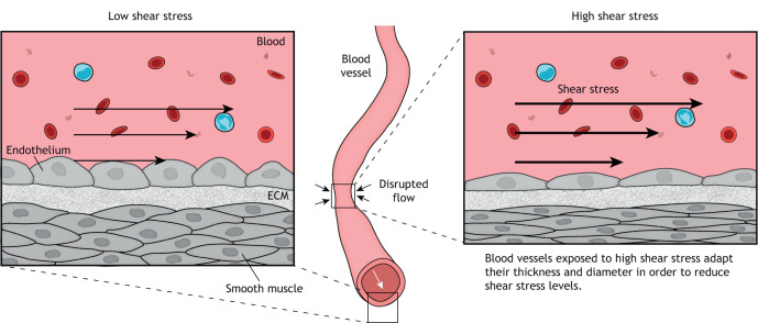
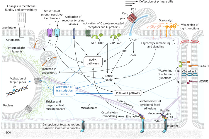
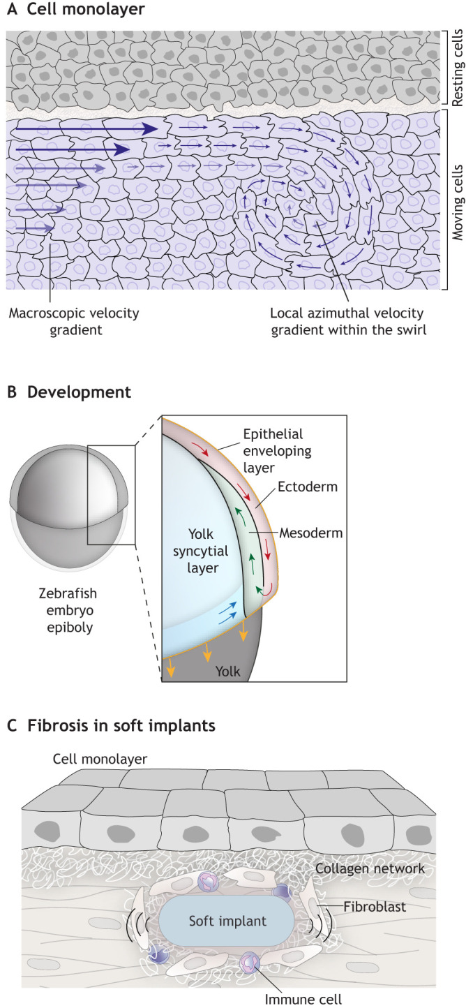
Shear stress is essential for normal physiology and malignancy. Common physiological processes - such as blood flow, particle flow in the gut, or contact between migratory cell clusters and their substrate - produce shear stress that can have an impact on the behavior of different tissues. In addition, shear stress has roles in processes of biomedical interest, such as wound healing, cancer and fibrosis induced by soft implants. Thus, understanding how cells react and adapt to shear stress is important. In this Review, we discuss in vivo and in vitro data obtained from vascular and epithelial models; highlight the insights these have afforded regarding the general mechanisms through which cells sense, transduce and respond to shear stress at the cellular levels; and outline how the changes cells experience in response to shear stress impact tissue organization. Finally, we discuss the role of shear stress in collective cell migration, which is only starting to be appreciated. We review our current understanding of the effects of shear stress in the context of embryo development, cancer and fibrosis, and invite the scientific community to further investigate the role of shear stress in these scenarios.

Keywords: Biomechanics; Cytoskeleton; Fluid shear stress; Shear stress.


Conflict of interest statement

Competing interests The authors declare no competing or financial interests.

Figures

Fig. 1.
Fig. 1.
Schematic illustration of how shear stress can affect blood vessels. Blood flow generates shear stress at the surface of the endothelial cells lining the blood vessel, to which the vessel responds to maintain normal shear stress levels. Under low physiological levels of shear stress, the vessel increases its thickness (left), specifically of the tunica media, and so decreases the internal diameter. This is achieved by thickening of the cellular body and loss of cell polarity. In areas of higher stress, such as in constrictions, the thickness of the vessel is reduced, and thus the internal diameter is increased (right). This occurs via a cellular mechanism involving elongation of cells and polarization of the cells in the direction of blood flow through activation of pathways that promote vasodilation.
Fig. 2.
Fig. 2.
Cellular responses to shear stress. Simplified representation of the molecular mechanisms involved in mechanosensing and mechanotransduction triggered in different cell compartments in response to shear stress. At the membrane, shear stress induces changes in fluidity and permeability, which can lead to activation of membrane-bound molecules, including activation of G proteins independently of the G-protein-coupled receptors. Membrane-bound receptors (such as receptor tyrosine kinases and G-protein-coupled receptors) and stretch-activated ion channels (such as PIEZO1) can also be activated by shear stress. FSS can bend primary cilia and trigger Ca2+ influx through activation of polycystin-1 (PC1) and polycystin-2 (PC2) transmembrane proteins. This rise in intracellular levels of Ca2+ can activate multiple signaling molecules and induce an increase in endocytosis of proteins and small molecules. Shear stress causes conformational changes in the glycocalyx, affecting the local concentration of molecules in the extracellular domain and inducing several signaling pathways in the intracellular domain, including those regulating the production of NO and the organization of the cytoskeleton. Activation of tyrosine kinases in FAs, such as FAK and Src, leads to activation of multiple signaling molecules and cytoskeleton reassembly in response to shear stress. FSS leads to a remodeling of FAs that is dependent on the stability of the actin stress fibers linked to them: disassembly of FAs anchored to inner perpendicular fibers is promoted, whereas FAs linked to peripheral fibers remain stable. Shear stress is associated with weakening of both tight junctions and adherent junctions. Adhesion proteins (such as PECAM-1 and VE-cadherin) in cell–cell junctions can sense and respond to shear stress, causing activation of VEGFR2 tyrosine kinase and induction of downstream signaling cascades such as the PI3K–AKT pathway. In response to shear stress, cytoskeletal fibers suffer major modifications and rearrangements. In the cytoplasm, mechanosensors induce activation of multiple signaling molecules and cascades, including PI3K–AKT and mitogen-activated protein kinase (MAPK) pathways. This causes several transcription factors (such as NRF2 and NF-κB) to be translocated to the nucleus, where they alter gene expression, leading to long-term cellular adaptations necessary to cope with different levels of shear stress. CaM, calmodulin; PKC, protein kinase C; Rho, Ras homolog proteins.
Fig. 3.
Fig. 3.
Shear stress in tissue contexts. (A) Diagram showing a top view of a cell monolayer sliding over a tissue. Migratory cells have a gradient of velocity from the border to the interior of the cluster. These gradients generate torsion of the velocity vector due to differential friction between migrating cells, in this way starting a swirl. In this context, the local azimuthal shear rate within the cell swirl is lower than the supracellular shear rate generated at the interface, which induces the swirl formation. (B) Schematic illustration of a side view of a zebrafish embryo during gastrulation, with an inset showing associated cell movement. In this case, shear stress is generated at the interface between ectoderm and mesoderm progenitors that migrate in opposite directions. These forces are required to determine the position of the neural progenitors. (C) Diagram showing how fibrosis develops from soft implants. Here, friction leading to shear stress can occur at the boundary between soft implants and surrounding tissues. This shear stress promotes inflammation and deposition of extracellular components in the surrounding connective tissue, leading to fibrosis.

References

    1. Aboualaiwi, W. A., Takahashi, M., Mell, B. R., Jones, T. J., Ratnam, S., Kolb, R. J. and Nauli, S. M. (2009). Ciliary polycystin-2 is a mechanosensitive calcium channel involved in nitric oxide signaling cascades. Circ. Res. 104, 860-869. 10.1161/CIRCRESAHA.108.192765 - DOI - PMC - PubMed
    1. Albuquerque, M. L., Waters, C. M., Savla, U., Schnaper, H. W. and Flozak, A. S. (2000). Shear stress enhances human endothelial cell wound closure in vitro. Am. J. Physiol. Heart Circ. Physiol. 279, H293-H302. 10.1152/ajpheart.2000.279.1.H293 - DOI - PubMed
    1. Avvisato, C. L., Yang, X., Shah, S., Hoxter, B., Li, W., Gaynor, R., Pestell, R., Tozeren, A. and Byers, S. W. (2007). Mechanical force modulates global gene expression and beta-catenin signaling in colon cancer cells. J. Cell Sci. 120, 2672-2682. 10.1242/jcs.03476 - DOI - PubMed
    1. Baeyens, N., Bandyopadhyay, C., Coon, B. G., Yun, S. and Schwartz, M. A. (2016). Endothelial fluid shear stress sensing in vascular health and disease. J. Clin. Invest. 126, 821-828. 10.1172/JCI83083 - DOI - PMC - PubMed
    1. Baghban, R., Roshangar, L., Jahanban-Esfahlan, R., Seidi, K., Ebrahimi-Kalan, A., Jaymand, M., Kolahian, S., Javaheri, T. and Zare, P. (2020). Tumor microenvironment complexity and therapeutic implications at a glance. Cell Commun. Signal 18, 59. 10.1186/s12964-020-0530-4 - DOI - PMC - PubMed

Publication types