Review
. 2023 Nov;83(16):1475-1499.
doi: 10.1007/s40265-023-01940-2. Epub 2023 Sep 25.

Current Understanding of Complement Proteins as Therapeutic Targets for the Treatment of Immunoglobulin A Nephropathy

Affiliations
Review

Current Understanding of Complement Proteins as Therapeutic Targets for the Treatment of Immunoglobulin A Nephropathy

Arun Rajasekaran et al. Drugs. 2023 Nov.

Abstract

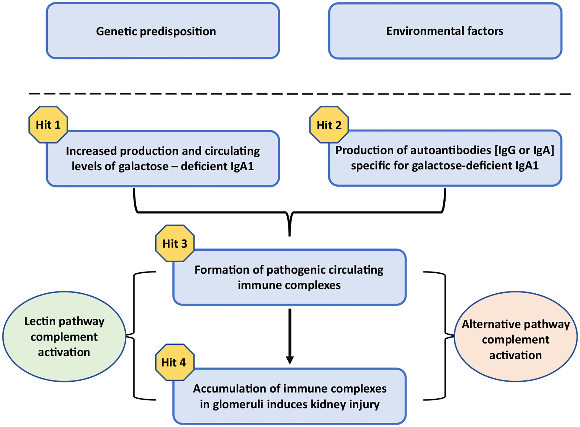
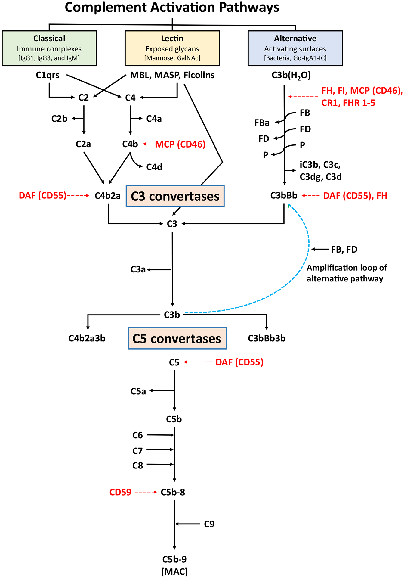
Immunoglobulin A nephropathy (IgAN) is the most common primary glomerulonephritis worldwide and a frequent cause of kidney failure. Currently, the diagnosis necessitates a kidney biopsy, with routine immunofluorescence microscopy revealing IgA as the dominant or co-dominant immunoglobulin in the glomerular immuno-deposits, often with IgG and sometimes IgM or both. Complement protein C3 is observed in most cases. IgAN leads to kidney failure in 20-40% of patients within 20 years of diagnosis and reduces average life expectancy by about 10 years. There is increasing clinical, biochemical, and genetic evidence that the complement system plays a paramount role in the pathogenesis of IgAN. The presence of C3 in the kidney immuno-deposits differentiates the diagnosis of IgAN from subclinical glomerular mesangial IgA deposition. Markers of complement activation via the lectin and alternative pathways in kidney-biopsy specimens are associated with disease activity and are predictive of poor outcome. Levels of select complement proteins in the circulation have also been assessed in patients with IgAN and found to be of prognostic value. Ongoing genetic studies have identified at least 30 loci associated with IgAN. Genes within some of these loci encode complement-system regulating proteins that can interact with immune complexes. The growing appreciation for the central role of complement components in IgAN pathogenesis highlighted these pathways as potential treatment targets and sparked great interest in pharmacological agents targeting the complement cascade for the treatment of IgAN, as evidenced by the plethora of ongoing clinical trials.


Conflict of interest statement

JN is a co-founder and co-owner of and consultant for Reliant Glycosciences, LLC. JN is a co-inventor on US patent application 14/318,082 (assigned to UAB Research Foundation). JN has a sponsored research agreement with Travere Therapeutics. BAJ is a co-founder and co-owner of Reliant Glycosciences, LLC and a co-inventor on US patent application 14/318,082 (assigned to UAB Research Foundation). MBR is a co-founder and co-owner of and consultant for Reliant Glycosciences, LLC. MBR is a co-inventor on US patent application 14/318,082 (assigned to UAB Research Foundation). DVR received research funding from Reata Pharmaceuticals, Travere Therapeutics (Retrophin), Pfizer Pharmaceuticals, Calliditas Therapeutics (Pharmalink), Otsuka Pharmaceuticals (Visterra), Vertex Pharmaceuticals, Chinook Pharmaceuticals, consultancy fees from Novartis, GSK, George Clinical, Eledon Pharmaceuticals, Otsuka Pharmaceuticals (Visterra), Calliditas Therapeutics (Pharmalink), Chinook Pharmaceuticals. DVR is co-founder and co-owner of Reliant Glycosciences, LLC. None of the listed commercial entities contributed to this study. AR and TJG have nothing to disclose.

Figures

Fig. 1
Fig. 1
Proposed four-hit hypothesis for pathogenesis of immunoglobulin A nephropathy (IgAN). In genetically susceptible individuals, environmental factors are thought to trigger the onset of IgAN. A four-hit hypothesis has been proposed for the disease pathogenesis: increased levels of circulatory galactose-deficient IgA1 (Hit 1) lead to production of autoantibodies (either IgG or IgA, but mostly of the IgG isotype) (Hit 2). This process results in the formation of circulating nephritogenic immune complexes (Hit 3), some of which deposit in the glomeruli and induce kidney injury via mesangial cell activation and proliferation (Hit 4) [32]. There is evidence that the alternative complement pathway and, at least in some patients, the lectin pathway are involved in the pathogenesis of IgAN
Fig. 2
Fig. 2
Simplified schema of the complement pathways. The three pathways of complement activation, classical, lectin, and alternative, are initiated by interactions of the complement proteins with distinct structures. Antigen-antibody complexes can activate the classical pathway. Mannose-binding lectin (MBL) recognizes carbohydrate structures, and upon association with serine proteases (MASP, mannose-associated serine proteases), can activate the lectin pathway. Complement C3 that is covalently bound to pathogen surfaces via a thioester bond as C3b initiates the alternative complement pathway. Each pathway ultimately generates an active C3 convertase, resulting in cleavage of C3 into C3a and C3b fragments. C3b can interact with C4b2a of the classical pathway or C3bBb of the alternative pathway to produce C5 convertase that cleaves C5 into C5a and C5b fragments. In the alternative complement pathway, every C3b molecule generated serves as the initiating point for a new C3 convertase (C3bBb), thereby generating numerous C3b molecules via the “amplification loop” process. In all complement pathways, C5b binds to the cell membrane of the pathogen and serves as a platform for assembly of the membrane attack complex (MAC). The formation of MAC can be inhibited by membrane-bound CD59. Several other regulatory proteins of the complement-activation pathways are shown in red. FB and FD perpetuate the alternative pathway “amplification loop”. CR1 complement receptor 1, DAF decay-accelerating factor, FB factor B, FD factor D, FHR 1–5 factor H-related proteins 1–5, FI factor I, GalNAc N-acetylgalactosamine, Gd-IgA1 galactose-deficient IgA1, Gd-IgA1-IC galactose-deficient IgA1-containing immune complexes, MCP membrane cofactor protein, P properdin
Fig. 3
Fig. 3
Structures of human complement C3 and C3 cleavage products and the thioester bond. IgA1-containing circulating immune complexes in IgAN patients have C3, presumably covalently attached through the thioester bond to IgA1 and/or IgG, as reported for other types of immune complexes [–116]. A Domain arrangement of C3 β and α chains. Domains are individually colored and labeled. Domain-starting residues are noted below the schematic. Residue numbering and colors follow those in the publication of Janssen et al [118]. B, C Surface rendering and cartoon models of C3 (PDB ID: 2A73 [118]) are shown. The conformation of C3 protects the thioester bond (red spheres boxed in [C]) from nucleophilic activation. DF Cleavage products of C3 are shown in surface representation. (D) C3b (PDB ID: 2I07 [190]) undergoes considerable conformational change, most notable is the movement of the thioester-containing domain (TED). This change exposes and activates residues C988 and Q991 (labeled and shaded in red on structures in DF), which are involved in formation of a thioester bond. E iC3b (PDB ID: 7AKK [191]) and F C3dg (derived from PDB ID: 7AKK) are shown. G A close-up view of the boxed area in C is shown with the thioester bond represented with stick model. The thioester bond in the structure of iC3b is broken and shown as the amino acid residues C988 and Q991. This view is from the boxed area in E. IgAN immunoglobulin A nephropathy, PDB ID Protein Data Bank identification code

References

    1. Berger J, Hinglais N. intercapillary deposits of IgA-IgG. J Urol Nephrol (Paris). 1968;74(9):694–5. - PubMed
    1. Berthoux FC, Mohey H, Afiani A. Natural history of primary IgA nephropathy. Semin Nephrol. 2008;28(1):4–9. 10.1016/j.semnephrol.2007.10.001. - DOI - PubMed
    1. Hastings MC, Bursac Z, Julian BA, Villa Baca E, Featherston J, Woodford SY, et al. Life expectancy for patients from the southeastern United States with IgA nephropathy. Kidney Int Rep. 2018;3(1):99–104. 10.1016/j.ekir.2017.08.008. - DOI - PMC - PubMed
    1. Jennette JC. The immunohistology of IgA nephropathy. Am J Kidney Dis. 1988;12(5):348–52. 10.1016/s0272-6386(88)80022-2. - DOI - PubMed
    1. Conley ME, Cooper MD, Michael AF. Selective deposition of immunoglobulin A1 in immunoglobulin A nephropathy, anaphylactoid purpura nephritis, and systemic lupus erythematosus. J Clin Invest. 1980;66(6):1432–6. 10.1172/jci109998. - DOI - PMC - PubMed

LinkOut - more resources