. 2023 Oct 11;480(19):1583-1598.
doi: 10.1042/BCJ20230267.

Proteotoxic stresses stimulate dissociation of UBL4A from the tail-anchored protein recognition complex

Affiliations

Proteotoxic stresses stimulate dissociation of UBL4A from the tail-anchored protein recognition complex

Takumi Hagiwara et al. Biochem J. .

Abstract

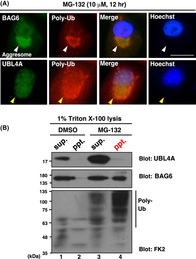
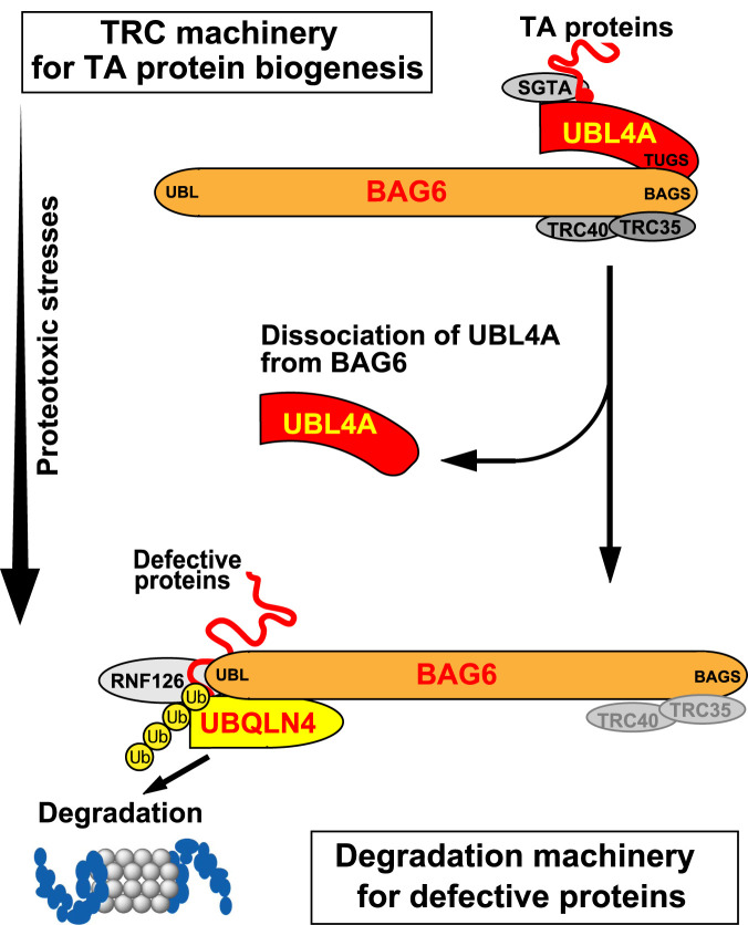
Inclusion body formation is associated with cytotoxicity in a number of neurodegenerative diseases. However, the molecular basis of the toxicity caused by the accumulation of aggregation-prone proteins remains controversial. In this study, we found that disease-associated inclusions induced by elongated polyglutamine chains disrupt the complex formation of BAG6 with UBL4A, a mammalian homologue of yeast Get5. UBL4A also dissociated from BAG6 in response to proteotoxic stresses such as proteasomal inhibition and mitochondrial depolarization. These findings imply that the cytotoxicity of pathological protein aggregates might be attributed in part to disruption of the BAG6-UBL4A complex that is required for the biogenesis of tail-anchored proteins.

Keywords: BAG6; UBL4A; polyglutamine disease; proteasome; protein quality control; tail-anchored protein.


Conflict of interest statement

The authors declare that there are no competing interests associated with the manuscript.

Figures

Figure 1.
Figure 1.. Schematic of the functional duality of BAG6 complex.
UBL4A, TRC35, and TRC40, mammalian homologues of yeast Get5, Get4, and Get3, respectively, are complexed with the C-terminus of BAG6 for the correct insertion of tail-anchored (TA) proteins into the lipid bilayer of the ER membrane (TA protein synthesis). In contrast, the N-terminal region of BAG6 is complexed with the ubiquitin ligase RNF126 [82] and UBQLN4, an ubiquitin receptor for 26S proteasomal targeting, and both of these are critical for the degradation of newly synthesized defective proteins. It has been reported that BAG6 tends to be enfolded into the aggresomes under proteasomal inhibition with polyubiquitinated aggregation-prone polypeptides. TMD, transmembrane domain; ER, endoplasmic reticulum.
Figure 2.
Figure 2.. UBL4A is absent from the MG-132-induced insoluble aggresome fraction.
(A) Immunosignals of BAG6 (green in upper panels) and polyubiquitin (red) were positive in insoluble aggresomes following MG-132 treatment, while those of UBL4A (green in lower panels) were negative. HeLa cells were treated with MG-132 (10 µM) for 12 h, and fixed cells were stained with anti-BAG6, anti-UBL4A, and anti-polyubiquitin antibodies. The positions of the aggresomes are indicated by arrowheads. Hoechst nuclear stains are merged in right panels. Bar: 10 µm. (B) FLAG-UBL4A-transfected HeLa cells were treated with 10 µM MG-132 or DMSO (as a negative control) for 6 h. Cellular homogenates with 1% Triton X-100 lysis buffer were fractionated by centrifugation at 20 000×g to obtain the supernatant (sup.) and pellet (ppt.) fractions, which were immunoblotted with anti-FLAG (UBL4A), anti-BAG6, and anti-polyubiquitin (FK2) antibodies.
Figure 3.
Figure 3.. Association of UBL4A and BAG6 is suppressed by proteasome inhibition in HeLa cells.
(A and B) MG-132 treatments reduced the amounts of BAG6-associated FLAG-UBL4A in soluble fractions of HeLa cells. Expression plasmids of FLAG-tagged UBL4A, TRC35, and TRC40 were transfected into HeLa cells. After 24 h of transfection, the cells were treated with (+) or without (−) 10 µM MG-132 for 4 h. Anti-BAG6 antibody co-precipitated UBL4A with endogenous BAG6 less efficiently in the presence of MG-132 (A, top panel), while the co-precipitation of TRC35 and TRC40 with BAG6 was not influenced by this treatment (B, top panel). Note that MG-132 treatment resulted in the accumulation of total soluble UBL4A protein, probably due to its stabilization (A, second panel). (C) MG-132-treatment of HeLa cells stimulated the association of UBQLN4 with BAG6. After 24 h of FLAG-UBQLN4 transfection, the cells were treated with (+) or without (−) 10 µM MG-132 for 4 h. Anti-BAG6 antibody co-precipitated UBQLN4 with endogenous BAG6 more efficiently in the presence of MG-132 (C, top panel), while the expression level of UBQLN4 was not affected by this treatment (C, middle panel). (D) Schematic image of mutually exclusive associations of UBQLN4 and UBL4A with BAG6 in cells treated with (+) or without (−) proteasome inhibitor MG-132.
Figure 4.
Figure 4.. Forced expression of UBL4A blocks the translocation of BAG6 to the insoluble fractions.
(A) Translocation of endogenous BAG6 to insoluble aggregates was suppressed by overexpression of UBL4A in proteasome-suppressed cells. After cells were treated with 5 µM MG-132 for 18 h, the cells were fractionated to soluble (sup.) and insoluble (ppt.) fractions by centrifugation at 10 000×g with buffer containing 1% Triton X-100. The resulting sup. and ppt. fractions were subjected to western blot analyses with anti-BAG6 and anti-FLAG (UBL4A) antibodies. Vimentin was used as a loading control and insoluble fraction marker. Note that increased amount of FLAG-UBL4A can be detected in the soluble fraction of MG-132-treated cells (lane 4), even though it was essentially negative in insoluble fractions (lane 8). (B) The graph indicates the quantified signal intensities of the endogenous BAG6 protein in insoluble fractions relative to the anti-vimentin signals. Experiments involved three biologically independent replicates to compute statistical significance. Data are presented as means ± standard deviation (S.D.) and were analyzed using Student's t-test. A P-value <0.05 was considered statistically significant.
Figure 5.
Figure 5.. Aggregation of expanded polyQ protein disrupts of the UBL4A–BAG6 association.
(A) FLAG-UBL4A was immunoprecipitated from polyQ-GFP-expressing HeLa cell extracts (5% input), and precipitates (IP: FLAG) were subjected to western blot analysis with an anti-BAG6 antibody to quantify the amount of endogenous BAG6 that was associated with FLAG-UBL4A (top panel). Expressions of polyQ proteins were verified by GFP blots (bottom panel). (B) Endogenous BAG6 was immunoprecipitated by anti-BAG6 (or non-immune control IgG) from the cell extracts of Q79-GFP (+) or mock (−)-expressing HeLa cells. Precipitates were subjected to western blot analysis with an anti-UBL4A antibody to quantify the amounts of BAG6-associated UBL4A protein (top panel). siRNA for UBL4A was performed (+) to confirm the validity of the UBL4A immunosignal. Anti-BAG6 and anti-Hsc70 blot verified the quantitative immunoprecipitation. The Hsc70 blot in the input panel shows the loading control. ‘Endo.' stands for endogenous proteins. (C) Quantitative evaluation of the endogenous BAG6 signal that co-precipitated with FLAG-UBL4A from polyQ-GFP-expressing cell extracts. Note that the value of co-precipitated BAG6 from cells not expressing polyQ protein (Q0) was used as the standard (100%). Experiments involved six biologically independent replicates to compute statistical significance. Data are presented as means ± standard deviation (S.D.) and were analyzed using Student's t-test. A P-value <0.05 was considered statistically significant. (D) Schematic of the NanoBiT split luciferase assay used in this study. Large-BiT (LgBiT, 17.6 kDa) and Small-BiT (SmBiT, 11 amino acids long) tags were fused to the C-termini of BAG6 and UBL4A, respectively, to detect the binding efficiency of these proteins in cells. Note that complex formation between BAG6–UBL4A is directly mediated through their C-terminal BAGS-TUGS domains [61,62]. The numbers denote the corresponding amino acid positions of full-length BAG6 protein. (E) PolyQ32 and polyQ79 proteins were expressed in HeLa cells with BAG6-LgBiT and UBL4A-SmBiT. Forty-eight hours after transfection of expression plasmids, NanoBiT-derived luminescence were measured, and the respective values normalized by cell number (RLU) were plotted on a graph. Welch's t-test n = 4. * P-value <0.05 was considered statistically significant.
Figure 6.
Figure 6.. Mitochondrial depolarization leads to dissociation of the UBL4A–BAG6 complex.
(A) Time-dependent dissociation of BAG6–UBL4A complex by treatment with CCCP, a mitochondrial uncoupling agent. Twenty-four hours after transfection with BAG6-LgBiT and UBL4A-SmBiT expression vectors, FLAG-Parkin-expressing HeLa cells were treated with 20 µM CCCP for the indicated time periods, and NanoBiT-derived luminescence was measured, and the respective values normalized by cell number (RLU) were plotted on a graph. Student's t-test, n = 3. ** P < 0.01. (B) Twenty-four hours after transfection with BAG6-LgBiT and UBL4A-SmBiT expression vectors, FLAG-Parkin-expressing HeLa cells were treated with 20 µM CCCP for 4 h. Four-hour treatment with dimethyl sulfoxide (DMSO, a solvent for CCCP) was used as a negative control. Welch's t-test, n = 4. ** P < 0.01.
Figure 7.
Figure 7.. Possible model for proteotoxic stress-induced defects in TRC machinery.
TRC machinery including BAG6–UBL4A complex participates in tail-anchored (TA) protein biogenesis. Once proteotoxic stresses (such as polyQ aggregation and mitochondrial depolarization) are induced, the BAG6–UBL4A complex dissociates, letting BAG6 separate from UBL4A. Because the UBL4A–BAG6 complex is a core of mammalian TRC machinery, its dissociation may result in the attenuation of tail-anchored protein synthesis, thereby leading to partial cytotoxicity. Note that many of these components are likely to form a dimer (or multimer), although they are described here as a monomer for simplicity.

Similar articles

References

    1. Ross, C.A. and Poirier, M.A. (2004) Protein aggregation and neurodegenerative disease. Nat. Med. 10, S10–S17 10.1038/nm1066 - DOI - PubMed
    1. Broadley, S.A. and Hartl, F.U. (2009) The role of molecular chaperones in human misfolding diseases. FEBS Lett. 583, 2647–2653 10.1016/j.febslet.2009.04.029 - DOI - PubMed
    1. Chakrabarti, O., Ashok, A. and Hegde, R.S. (2009) Prion protein biosynthesis and its emerging role in neurodegeneration. Trends Biochem. Sci. 34, 287–295 10.1016/j.tibs.2009.03.001 - DOI - PMC - PubMed
    1. Schezinger, E., Lurz, R., Turmaine, M., Mangiarini, L., Hollenbach, B., Hasenbank, R.et al. (1997) Huntingtin-encoded polyglutamine expansions form amyloid-like protein aggregates in vitro and in vivo. Cell 90, 549–558 10.1016/s0092-8674(00)80514-0 - DOI - PubMed
    1. Igarashi, S., Koide, R., Shimohata, T., Yamada, M., Hayashi, Y., Takano, H.et al. (1998) Suppression of aggregate formation and apoptosis by transglutaminase inhibitors in cells expressing truncated DRPLA protein with an expanded polyglutamine stretch. Nat. Genet. 18, 111–117 10.1038/ng0298-111 - DOI - PubMed