Observational Study
. 2023 Nov 1;43(11):1890-1903.
doi: 10.1097/IAE.0000000000003927. Epub 2023 Oct 19.

MULTIZONAL OUTER RETINOPATHY AND RETINAL PIGMENT EPITHELIOPATHY (MORR): A Newly Recognized Entity or an Unusual Variant of AZOOR?

Affiliations
Observational Study

MULTIZONAL OUTER RETINOPATHY AND RETINAL PIGMENT EPITHELIOPATHY (MORR): A Newly Recognized Entity or an Unusual Variant of AZOOR?

Prithvi Ramtohul et al. Retina. .

Abstract

Purpose: To describe specific clinical, multimodal imaging, and natural history features of an unusual variant of acute zonal occult outer retinopathy.

Methods: Retrospective, observational, longitudinal, multicenter case series. Patients exhibiting this unusual clinical condition among cases previously diagnosed with acute zonal occult outer retinopathy were included. Multimodal imaging, laboratory evaluations, and genetic testing for inherited retinal diseases were reviewed.

Results: Twenty eyes from 10 patients (8 females and 2 males) with a mean age of 54.1 ± 13.3 years (range, 38-71 years) were included. The mean follow-up duration was 13.1 ± 5.3 years (range, 8-23 years). Presenting symptoms were bilateral in 7 patients (85% of eyes) and included scotomata and photopsia. All patients had bilateral lesions at presentation involving the peripapillary and far peripheral retina. Baseline optical coherence tomography showed alteration of the retinal pigment epithelium and photoreceptor layers corresponding to zonal areas of fundus autofluorescence abnormalities. Centrifugal and centripetal progression of the peripapillary and far-peripheral lesions, respectively, occurred over the follow-up, resulting in areas of complete outer retinal and retinal pigment epithelium atrophy.

Conclusion: Initial alteration of photoreceptors and retinal pigment epithelium and a stereotypical natural course that includes involvement of the far retinal periphery, characterize this unusual condition. It may represent a variant of acute zonal occult outer retinopathy or may be a new entity. We suggest to call it multizonal outer retinopathy and retinal pigment epitheliopathy .


Conflict of interest statement

K. B. Freund is a consultant for Heidelberg Engineering, Zeiss, Allergan, Bayer, Genentech, and Novartis and receives research support from Genentech/Roche. None of the remaining authors has any financial/conflicting interests to disclose.

Figures

Fig. 1.
Fig. 1.
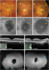
Multimodal imaging correlation of initial stage and long-term course of MORR (Case 8). A. At baseline, color fundus photography of the right eye shows a peripapillary lesion characterized by a well-demarcated yellow–gray core of RPE alterations (yellow arrowhead) and bordered by a thin hyperpigmented demarcation line (white arrowhead). B. At baseline, FAF image of the right eye shows the peripapillary lesion characterized by a speckled hyperautofluorescent core (yellow arrowhead) surrounded by a thin, continuous, hyperautofluorescent demarcation line (white arrowhead). C. At baseline, intermediate phase of fluorescein angiography of the right eye shows a hyperfluorescent peripapillary core (window defect, yellow arrowhead) and blockage of the pigmented demarcation line (white arrowhead). Note the absence of optic disk or retinal vascular staining or leakage. D. At baseline, late phase of indocyanine green angiography of the right eye shows hypofluorescence of the peripapillary core (reduced RPE uptake, yellow arrowhead). The demarcation line shows no distinctive features on indocyanine green angiography (white arrowhead). E and F. At baseline, near infrared reflectance image (E) of the right eye shows the peripapillary lesion with hyperreflective changes within the demarcation line (white arrowhead). The green line indicates the location of the OCT B-scan displayed in (F). Corresponding OCT B-scan (F) shows RPE disruption at the core, including RPE thickening interspersed with focal RPE atrophy (between white arrowheads). The hyperautofluorescent demarcation line colocalizes with focal RPE mottling (white arrowheads). The ellipsoid zone is attenuated, but still visible above areas of RPE alterations. G–J. Fundus autofluorescence images of the right eye acquired at 4 years (G), 4.5 years (H), 8 years (I), and 10 years (J). The peripapillary lesion shows centrifugal extension of the hypoautofluorescent core and shifting of the demarcation line toward the periphery. Note the episodic pattern of progression. The timepoint is displayed.
Fig. 2.
Fig. 2.
Peripapillary and far-peripheral lesions of initial stage of MORR (Case 9). A. At baseline, color fundus photography of the right eye shows a subtle peripapillary gray lesion of the RPE (white arrowhead). The timepoint is displayed. B. At baseline, color fundus photography of the left eye shows a more apparent peripapillary gray lesion of the RPE surrounded by an orange demarcation line (white arrowheads). The timepoint is displayed. C. At baseline, FAF image of the right eye shows a subtle peripapillary lesion with hyperautofluorescent features (white arrowhead). The timepoint is displayed. D. At baseline, FAF image of the left eye shows a peripapillary lesion with a speckled hyperautofluorescent core surrounded by a thin continuous hyperautofluorescent demarcation line (white arrowhead). The timepoint is displayed. E and F. At 2 years, FAF images of the right (E) and left (F) eye show centrifugal progression of the peripapillary lesion characterized by a hypoautofluorescent core surrounded by a large interrupted demarcation line with fringe-like hyperautofluorescent features radiating outward (white arrowheads). The timepoint is displayed. G and H. At 2 years, ultra-widefield pseudocolor fundus photographs of the right (G) and left (H) eye show far-peripheral lesions characterized by well-demarcated, 360-degree, annular zones of RPE atrophy accompanied by large spots of RPE hyperpigmentation (blue arrowheads). The timepoint is displayed. I and J. At 2 years, ultra-widefield FAF images of the right (I) and left (J) eye show peripapillary lesions (white arrowheads) and the far-peripheral annular lesions (blue arrowheads). The timepoint is displayed. K and L. At 2 years, horizontal OCT B-scans through the fovea of the right (K) and left eye (L) show peripapillary RPE disruption and attenuation of the ellipsoid zone (white arrowheads). The timepoint is displayed.
Fig. 3.
Fig. 3.
Initial stage and 15-year follow-up multimodal imaging of MORR (Case 1). A. At presentation, color fundus photography of the right eye acquired few days after symptoms' onset shows a well-demarcated yellow–gray core of RPE alterations (yellow arrowhead) bordered by a thin, yellowish, drusen-like demarcation line (white arrowhead). Note the absence of optic disk edema, perivascular exudation or sheathing, or vitritis. The timepoint is displayed. B. At 6 years, the size of the peripapillary lesion (yellow arrowhead) is relatively stable and attenuation of the demarcation line is noted (white arrowhead). The timepoint is displayed. C. At 15 years, centrifugal progression of the peripapillary lesion is noted. The core (yellow arrowhead) shows extension of the RPE atrophy and increased visibility of the choroidal vasculature. The demarcation line is shifted toward the periphery (white arrowhead). The timepoint is displayed. D. At presentation, FAF image shows a speckled hyperautofluorescent core (yellow arrowhead) surrounded by a thin, continuous, hyperautofluorescent demarcation line (white arrowhead). The timepoint is displayed. E. At 6 years, FAF image shows extension of the hypoautofluorescent core (yellow arrowhead), which is more apparent than seen on color fundus photography. The pattern of the demarcation line progresses into a larger interrupted border with fringe-like hyperautofluorescent features radiating outward (white arrowhead). The timepoint is displayed. F. At 15 years, centrifugal extension of the hypoautofluorescent core (yellow arrowhead) is noted. The demarcation line is shifted toward the periphery (white arrowhead). Note the thinning of the demarcation line and the reduced amount of the hyperautofluorescent features radiating outward. The timepoint is displayed. G. At 6 years, OCT B-scan through the fovea shows subtle RPE alterations in the peripapillary area (white arrowhead). The overlying ellipsoid zone is attenuated. The inset is the near infrared reflectance image with the green line indicating the location of the OCT B-scan. The timepoint is displayed. H. At 15 years, tracked OCT B-scan shows progression of the RPE atrophy toward the fovea (white arrowhead). Note the loss of the overlying ellipsoid zone and outer nuclear layer, and the secondary thinning of the underlying choroid. The inset is the near infrared reflectance image with the green line indicating the location of the OCT B-scan. The timepoint is displayed. I and J. At 15 years, ultra-widefield fundus autofluorescence images of the right (I) and left eye (J) show bilateral intermediate-stage peripapillary lesions (white arrowheads). Note the far-peripheral, annular, hypoautofluorescent lesions bordered by an interrupted demarcation line with fringe-like hyperautofluorescent features radiating inward (blue arrowheads). The timepoint is displayed.
Fig. 4.
Fig. 4.
Complete RPE degeneration resulting from merging of peripapillary and far-peripheral lesions in MORR (Case 4). A. At baseline, color fundus photography of the right eye shows peripapillary RPE alterations involving the fovea (white arrowhead). The timepoint is displayed. B. At baseline, color fundus photography of the left eye shows a well-demarcated, peripapillary yellowish–gray lesion surrounded by an orange demarcation line (white arrowhead). The timepoint is displayed. C. At 3 years, montage of FAF images of the right eye shows a peripapillary lesion with a hypoautofluorescent core and surrounded by large interrupted demarcation line with fringe-like hyperautofluorescent features radiating outward (white arrowhead). The timepoint is displayed. D. At 3 years, montage of FAF images of the left eye shows progression of the peripapillary lesion with a hypoautofluorescent core and surrounded by large interrupted demarcation line with fringe-like hyperautofluorescent features radiating outward (white arrowhead). Note the far-peripheral lesions inferiorly bordered by a large interrupted demarcation line with fringe-like hyperautofluorescent features radiating inward (blue arrowheads). The timepoint is displayed. E–J. Ultra-widefield FAF images of the right and left eye, respectively, acquired at 10 years (E and F), 13 years (G and H), and 21 years (I and J). The peripapillary lesion (white arrowheads) shows centrifugal extension of the hypoautofluorescent core and shifting of the demarcation line toward the periphery. Centripetal progression of the far-peripheral lesions is associated with merging of peripapillary and far-peripheral lesions, resulting in complete outer retinal and RPE degeneration at the final visit. The timepoint is displayed.
Fig. 5.
Fig. 5.
Episodic pattern of progression of MORR (Case 2). A. At baseline, ultra-widefield FAF image of the left eye acquired few days after symptoms' onset shows a subtle hyperautofluorescent peripapillary lesion (white arrowhead) and a macular lesion (green arrowhead). The timepoint is displayed. B. At 6 weeks, merging of the peripapillary and macular lesion is noted. The core of the peripapillary lesion shows a speckled hyperautofluorescence surrounded by a thin, continuous, hyperautofluorescent demarcation line (white arrowhead). Note the midperipheral lesions in the supero-temporal and infero-nasal areas (green arrowheads). The timepoint is displayed. C. At 6 years, ultra-widefield FAF image shows extension of the hypoautofluorescent core and minimal shifting of the demarcation line toward the periphery (white arrowhead). The pattern of the demarcation line progresses into a larger interrupted border with fringe-like hyperautofluorescent features radiating outward (white arrowhead). Note the centripetal progression of the hypoautofluorescent far-peripheral lesions (blue arrowheads). The timepoint is displayed. D. At 13 years, ultra-widefield FAF image shows further extension of the hypoautofluorescent core and minimal shifting of the demarcation line toward the periphery (white arrowhead). Attenuation and thinning of the demarcation line with loss of the hyperautofluorescent features are observed (white arrowhead). Note the relative stability of the hypoautofluorescent far-peripheral lesions (blue arrowheads). The timepoint is displayed. E. At 6 years, ultra-widefield FAF image of the right eye shows a peripapillary lesion with a hypoautofluorescent core surrounded by a large interrupted demarcation line with fringe-like hyperautofluorescent features radiating outward (white arrowhead). Note the annular hypoautofluorescent far-peripheral lesions (blue arrowhead). The timepoint is displayed. F. At 13 years, ultra-widefield FAF image of the right eye shows extension of the hypoautofluorescent core, thinning of the demarcation line with attenuation of the hyperautofluorescent features (white arrowhead). Note the centripetal progression of the hypoautofluorescent far-peripheral lesions (blue arrowhead). The timepoint is displayed. G and H. At 13 years, ultra-widefield pseudocolor fundus photographs of the right (G) and left eye (H) show peripapillary atrophy of the RPE and increased visibility of the choroidal vasculature (white arrowheads). Note the far-peripheral pigmented lesions (blue arrowheads). The timepoint is displayed. I. At 6 years, near infrared reflectance image of the right eye shows hyperreflective changes within the demarcation line radiating outward. The green line indicates the location of the OCT B-scan displayed in (J).J.At 6 years, OCT B-scan of the right eye shows peripapillary RPE disruption and loss of the ellipsoid zone and interdigitation zone. The hyperautofluorescent features of the demarcation line colocalizes with the focal RPE thickening (white arrowhead). The timepoint is displayed.K. At 13 years, near infrared reflectance image of the right eye shows centrifugal extension of the peripapillary lesion. The green line indicates the location of the OCT B-scan displayed in (L).L. At 13 years, OCT B-scan of the right eye shows extension of the peripapillary RPE disruption toward the fovea (white arrowhead). Note the further loss of the outer nuclear layer indicating photoreceptor cell loss and development of outer retinal tubulation (orange arrowhead). The timepoint is displayed.
Fig. 6.
Fig. 6.
Centrifugal and centripetal progression in MORR (Case 5). A–D. Ultra-widefield pseudocolor fundus photographs of the right eye acquired at baseline (A), 4 years (B), 6 years (C), and 10 years (D). At baseline (A), peripapillary (white arrowhead) and far-peripheral annular lesions of the RPE (blue arrowheads) are observed. Note the macular lesion (green arrowhead). During the follow-up, the peripapillary lesion (white arrowheads) shows centrifugal progression and merges with the macular lesion and the far-peripheral lesion at 6 and 10 years, respectively. Far-peripheral lesions are characterized by well-demarcated, 360-degree, annular zones of RPE atrophy accompanied by large spots of RPE hyperpigmentation (blue arrowheads). Centripetal progression of the far-peripheral lesions is noted. The timepoint is displayed. E–H. Ultra-widefield FAF images of the right eye acquired at baseline (E), 4 years (F), 6 years (G), and 10 years (H). At baseline (E), the peripapillary lesion shows a hypoautofluorescent core bordered by a large interrupted demarcation line with fringe-like hyperautofluorescent features radiating outward (white arrowhead). Note the macular lesion showing similar autofluorescent characteristics (green arrowhead). Far-peripheral annular lesions are hypoautofluorescent and bordered by a large interrupted demarcation line with fringe-like hyperautofluorescent features radiating inward (blue arrowheads). During the follow-up, the peripapillary lesion (white arrowheads) shows centrifugal extension of the hypoautofluorescent core and shifting of the demarcation line toward the periphery. Centripetal progression of the far-peripheral lesions associated with thinning and loss of the hyperautofluorescent features inside the demarcation line is noted at the final visit (blue arrowheads). The timepoint is displayed. I–L. Ultra-widefield pseudocolor fundus photographs of the left eye acquired at baseline (I), 4 years (J), 6 years (K), and 10 years (L). Centrifugal and centripetal progression of the peripapillary (white arrowhead) and far-peripheral (blue arrowheads) lesions, respectively, is noted during the follow-up. The timepoint is displayed. M–P. Ultra-widefield FAF images of the left eye acquired at baseline (M), 4 years (N), 6 years (O), and 10 years (P). The peripapillary lesion (white arrowheads) shows centrifugal extension of the hypoautofluorescent core and shifting of the demarcation line toward the periphery. Centripetal progression of the far-peripheral lesions associated with thinning and loss of the hyperautofluorescent features inside the demarcation line is noted at the final visit (blue arrowheads). The timepoint is displayed.
Fig. 7.
Fig. 7.
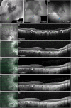
Fundus autofluorescence imaging and OCT of initial stage of MORR (Case 3). A. At baseline, montage of FAF images of the right eye shows a peripapillary lesion with a speckled hyperautofluorescent core superiorly and bordered by a thin continuous hyperautofluorescent demarcation line (white arrowhead). Note the midperipheral lesion inferiorly (green arrowhead). The timepoint is displayed. B. At 4 years, the peripapillary lesion shows centrifugal progression of the hypoautofluorescent core and shifting of the demarcation line toward the periphery (white arrowhead). Note the merging of the midperipheral lesion with far-peripheral lesions (blue arrowhead). The timepoint is displayed. C. At 10 years, the peripapillary lesion shows further centrifugal progression of the hypoautofluorescent core and shifting of the demarcation line toward the periphery (white arrowhead). Note the persistence of fringe-like hyperautofluorescent features inside the demarcation line and radiating outward. Centripetal progression of far-peripheral lesions is seen (blue arrowhead). The timepoint is displayed. D. At baseline, near infrared reflectance image of the right eye shows speckled hyperreflectivity of the early-stage peripapillary lesion. The green line indicates the location of the OCT B-scan shown in (E). E. At baseline, OCT B-scan of the core shows focal mottling of the RPE interspersed with RPE atrophy (between white arrowheads). Note the disruption of the overlying ellipsoid zone. The hyperautofluorescent demarcation line colocalizes with focal RPE mottling (white arrowheads). The timepoint is displayed. F–M. Near infrared reflectance images and corresponding tracked OCT B-scans acquired at 4 years (F and G), 5 years (H and I), 6 years (J and K), and 10 years (L and M). On OCT, the demarcation line (white arrowheads) shows progressive shifting toward the periphery and attenuation of the RPE thickening. Extension of the RPE atrophy at the core of the peripapillary lesion is associated with secondary loss of the overlying ellipsoid zone and outer nuclear layer and thinning of the underlying choroid. The timepoint is displayed.

Similar articles

Cited by

References

    1. Gass JD. Acute zonal occult outer retinopathy. Donders lecture: The Netherlands ophthalmological society, Maastricht, Holland, June 19, 1992. J Clin Neuroophthalmol 1993;13:79–97. - PubMed
    1. Gass JD, Agarwal A, Scott IU. Acute zonal occult outer retinopathy: a long-term follow-up study. Am J Ophthalmol 2002;134:329–339. - PubMed
    1. Mrejen S, Khan S, Gallego-Pinazo R, et al. . Acute zonal occult outer retinopathy: a classification based on multimodal imaging. JAMA Ophthalmol 2014;132:1089–1098. - PubMed
    1. Spaide RF. Collateral damage in acute zonal occult outer retinopathy. Am J Ophthalmol 2004;138:887–889. - PubMed
    1. Vadboncoeur J, Jampol LM, Goldstein DA. Acute zonal occult outer retinopathy: a case report of regression after an intravitreal dexamethasone (OZURDEX) implant. Retin Cases Brief Rep 2022;16:466–469. - PubMed

Publication types

Substances

Supplementary concepts