Review
. 2024 Jun;50(4):537-551.
doi: 10.1055/s-0043-1774796. Epub 2023 Sep 25.

A Perspective on How Fibrinaloid Microclots and Platelet Pathology May be Applied in Clinical Investigations

Affiliations
Review

A Perspective on How Fibrinaloid Microclots and Platelet Pathology May be Applied in Clinical Investigations

Etheresia Pretorius et al. Semin Thromb Hemost. 2024 Jun.

Abstract

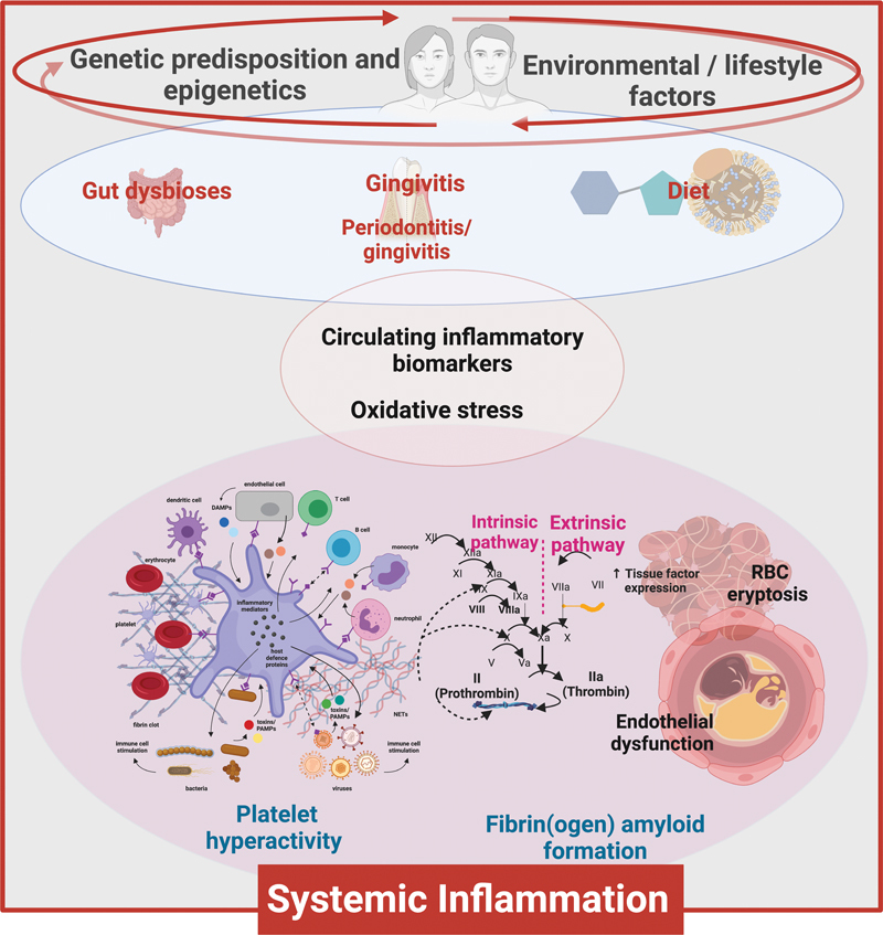
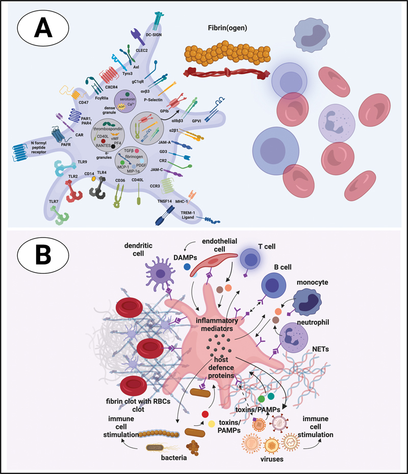
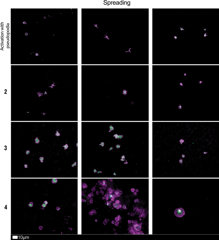
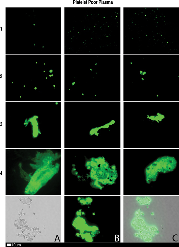
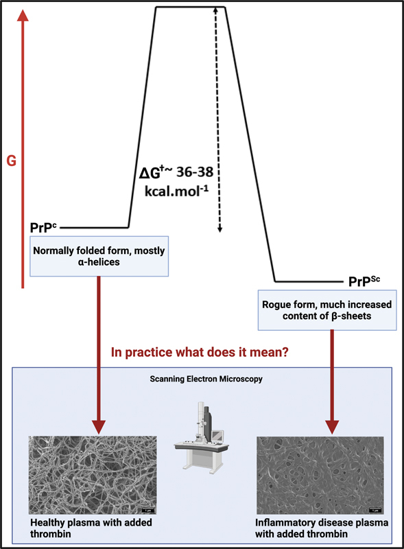
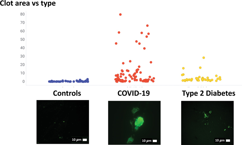
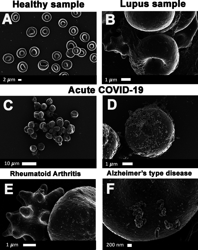
Microscopy imaging has enabled us to establish the presence of fibrin(ogen) amyloid (fibrinaloid) microclots in a range of chronic, inflammatory diseases. Microclots may also be induced by a variety of purified substances, often at very low concentrations. These molecules include bacterial inflammagens, serum amyloid A, and the S1 spike protein of severe acute respiratory syndrome coronavirus 2. Here, we explore which of the properties of these microclots might be used to contribute to differential clinical diagnoses and prognoses of the various diseases with which they may be associated. Such properties include distributions in their size and number before and after the addition of exogenous thrombin, their spectral properties, the diameter of the fibers of which they are made, their resistance to proteolysis by various proteases, their cross-seeding ability, and the concentration dependence of their ability to bind small molecules including fluorogenic amyloid stains. Measuring these microclot parameters, together with microscopy imaging itself, along with methodologies like proteomics and imaging flow cytometry, as well as more conventional assays such as those for cytokines, might open up the possibility of a much finer use of these microclot properties in generative methods for a future where personalized medicine will be standard procedures in all clotting pathology disease diagnoses.


Conflict of interest statement

Disclosure The authors report no conflicts of interest in this work.

Figures

Fig. 1
Fig. 1
Visualizing various factors that influence disease to the understand the phenomena of the disease due to the response to an external stimulus such as an infection. Figure created by authors using Biorender.com.
Fig. 2
Fig. 2
Platelet receptors and interactions with cells and proteins in circulation. Figure created by authors using Biorender.com.
Fig. 3
Fig. 3
Fluorescence microscopy imaging examples of the different stages of platelet activation and spreading that was used to score platelet activation in long COVID patients. After centrifuging freshly collected samples, the hematocrit fraction of each sample was retained and incubated for 30 minutes at room temperature with the two fluorescent markers, CD62P (PE-conjugated) (platelet surface P-selectin) (IM1759U, Beckman Coulter, Brea, CA) and PAC-1 (FITC-conjugated) (340507, BD Biosciences, San Jose, CA). 10 mL of each exposed sample was placed on microscope slide and viewed using a 63x oil objective. Stage 1, with minimally activated platelets, seen as small round platelets with a few pseudopodia, seen as healthy/control platelets that progresses to Stage 4, with egg-shaped platelets, indicative of spreading and the beginning of clumping (with permission from the CC-BY publication 28 ).
Fig. 4
Fig. 4
Fluorescence microscopy showing microclots in platelet poor plasma (PPP) with representative examples of the different stages of different stages of microclot formation. Stored or freshly prepared platelet poor plasma samples were exposed to Thioflavin T (ThT), a fluorogenic dye that binds to amyloid protein. A final concentration of 0.005 mM was used (Sigma-Aldrich, St. Louis, MO). Plasma was exposed for 30 minutes (protected from light) at room temperature, whereafter 3 µL stained PPP was placed on a glass slide and covered with a coverslip. Stage 1 shows minimal microclot formation in healthy/control PPP which progresses to the presence of the severe microclotting Stage 4. Bottom row represents examples of stage 4 microclots using ( A ) bright-field microscopy, ( B ) fluorescence microscopy, and ( C ) an overlay of fluorescence and bright-field microscopy (with permission from the CC-BY publication 28 ).
Fig. 5
Fig. 5
Protein–protein interactions may result in protein misfolding and have been shown to cause amyloidogenic changes to all kinds of proteins. This is illustrated in the upper part for the Prion protein PrP. The lower part shows electron micrographs of fibrin clots. Adapted in part from the CC-BY publications. We also note that the spike protein is itself amyloidogenic. Figure created by authors using Biorender.com.
Fig. 6
Fig. 6
Comparison of microclot area in healthy participants, and participants with Type 2 diabetes and acute COVID-19. (Raw data reanalyzed, and available in 26 ).
Fig. 7
Fig. 7
( A to F ) Whole blood scanning electron microscopy of micrograph data previously collected in published studies. ( A ) A few plasma deposits are discernible within the blood sample of a healthy participant ; ( B ) systemic lupus erythematosus ; ( C and D ) acute COVID-19 during the initial wave in 2020 ; ( E ) rheumatoid Arthritis ; ( F ) Alzheimer's-type disease.

Similar articles

Cited by

References

    1. Weisel J W. Fibrinogen and fibrin. Adv Protein Chem. 2005;70:247–299. - PubMed
    1. Adams R L, Bird R J. Review article: coagulation cascade and therapeutics update: relevance to nephrology. Part 1: overview of coagulation, thrombophilias and history of anticoagulants. Nephrology (Carlton) 2009;14(05):462–470. - PubMed
    1. Undas A, Ariëns R AS. Fibrin clot structure and function: a role in the pathophysiology of arterial and venous thromboembolic diseases. Arterioscler Thromb Vasc Biol. 2011;31(12):e88–e99. - PubMed
    1. Wolberg A S. Thrombin generation and fibrin clot structure. Blood Rev. 2007;21(03):131–142. - PubMed
    1. Palta S, Saroa R, Palta A. Overview of the coagulation system. Indian J Anaesth. 2014;58(05):515–523. - PMC - PubMed