. 2023 Sep 25;6(12):e202302122.
doi: 10.26508/lsa.202302122. Print 2023 Dec.

Two domains of Tim50 coordinate translocation of proteins across the two mitochondrial membranes

Affiliations

Two domains of Tim50 coordinate translocation of proteins across the two mitochondrial membranes

Marcel G Genge et al. Life Sci Alliance. .

Abstract

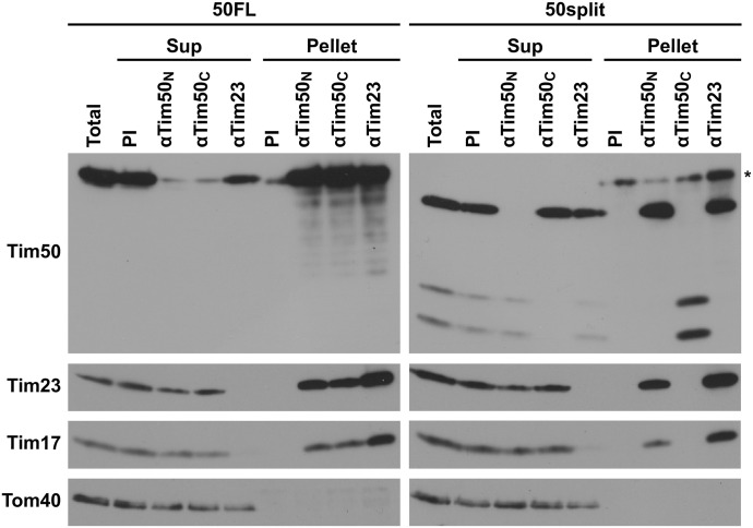
Hundreds of mitochondrial proteins with N-terminal presequences are translocated across the outer and inner mitochondrial membranes via the TOM and TIM23 complexes, respectively. How translocation of proteins across two mitochondrial membranes is coordinated is largely unknown. Here, we show that the two domains of Tim50 in the intermembrane space, named core and PBD, both have essential roles in this process. Building upon the surprising observation that the two domains of Tim50 can complement each other in trans, we establish that the core domain contains the main presequence-binding site and serves as the main recruitment point to the TIM23 complex. On the other hand, the PBD plays, directly or indirectly, a critical role in cooperation of the TOM and TIM23 complexes and supports the receptor function of Tim50. Thus, the two domains of Tim50 both have essential but distinct roles and together coordinate translocation of proteins across two mitochondrial membranes.


Conflict of interest statement

The authors declare that they have no conflict of interest.

Figures

Figure 1.
Figure 1.. Both domains of Tim50 in the IMS are essential for cell viability.
(A, C) Schematic representation of the domain structure of Tim50 and the variants analysed. 1 and 476 indicate the first and the last amino acid residues of Tim50 precursor, 131 indicates the start of the IMS segment, and 365 indicates the last residue of the core domain. TM, transmembrane segment; PBD, presequence-binding domain; IMS, intermembrane space; IM, inner membrane. (B, D) A Tim50 shuffling strain was transformed with centromeric plasmids encoding indicated variants of Tim50 under control of the endogenous promoter and 3′UTR. The ability of the Tim50 variants to support the function of the full-length protein was analysed on plates containing 5-FOA. Empty plasmid and a plasmid encoding a wild-type copy of Tim50 were used as negative and positive controls, respectively.
Figure 2.
Figure 2.. The function of Tim50 can be rescued by its two domains, core and PBD, expressed in trans.
(A) Schematic representation of the Tim50 variants expressed in trans. (B, C) A Tim50 shuffling strain was transformed with centromeric plasmids carrying the indicated Tim50 variants under control of endogenous promoter and 3′UTR. The ability of the Tim50 variants, alone or upon co-expression, to support the function of the full-length protein was analysed on plates containing 5-FOA at 24°C (B) or at 30°C (C). Empty plasmid and a plasmid encoding a WT copy of Tim50 were used as negative and positive controls, respectively. (D) Growth of indicated yeast strains was analysed by 10-fold serial dilution spot assay on plates containing rich medium with glucose (YPD) or lactate (YPLac), as fermentable and non-fermentable carbon sources, respectively. (E) Isolated mitochondria (10 and 20 μg) from 50FL and 50split cells were analysed by SDS–PAGE, followed by immunoblotting against depicted mitochondrial proteins. For simplicity reasons, the co-expression strain Tim50(1–370) + Tim50(366–476) was named “50split” and the corresponding wild-type strain expressing the full-length version of Tim50, Tim50(1–476), “50FL.”
Figure S1.
Figure S1.. Anchoring the core domain of Tim50 to the IM is crucial to rescue the function of Tim50, when co-expressed with a soluble PBD.
(A, C, E) Schematic representation of Tim50 and its variants expressed in trans. (B, D, F) A Tim50 shuffling strain was transformed with centromeric plasmids carrying indicated protein variants of Tim50 under control of endogenous promoter and 3′UTR. The ability of the individual or co-expressed Tim50 protein variants to support the function of the full-length protein was analysed on plates containing 5-FOA. Empty plasmid and a plasmid encoding a wild-type copy of Tim50 were used as negative and positive controls, respectively.
Figure 3.
Figure 3.. Tim50 is recruited to the TIM23 complex mainly through its core domain.
Isolated mitochondria from 50FL and 50split cells were solubilized with digitonin-containing buffer and subjected to immunoprecipitation with affinity-purified antibodies against Tim50N, Tim50C, and Tim23 prebound to Protein A Sepharose beads. Antibodies from pre-immune serum (PI) were used as a negative control. After washing, specifically bound proteins were eluted with Laemmli buffer. Total (20%), supernatant (Sup, 20%), and bound (Pellet, 100%) fractions were analysed by SDS–PAGE and immunoblotting with indicated antibodies. (*) indicates the heavy chains of the IgGs.
Figure 4.
Figure 4.. Protein import via the TIM23 complex and binding of Tim50 to precursors is impaired in 50split cells.
(A, B, C, D, E, F, G, H) 35S-labelled mitochondrial precursor proteins were imported into the mitochondria isolated from 50FL and 50split cells. After indicated time periods, aliquots were taken, import was stopped, and Proteinase K (PK) was added, where indicated. Mitochondria were reisolated and analysed by SDS–PAGE and autoradiography (upper panels). Quantifications of PK-protected mature forms of imported proteins are shown in the lower panels. The amount of the PK-protected mature form of imported proteins in the longest time point in 50FL mitochondria was set to 100%. Precursor (p), intermediate (i), and mature (m) forms of imported proteins. (*) indicates translation products synthesized from an internal methionine. (I) 35S-labelled Oxa1 precursor was imported into isolated 50FL and 50split mitochondria in the absence of membrane potential. Samples were subjected to cross-linking with 1,5-difluor-2,4-dinitrobenzol (DFDNB). After quenching of excess cross-linker, mitochondria were reisolated and solubilized in SDS-containing buffer to dissociate all noncovalent interactions. Samples were diluted in Triton X-100-containing buffer and subjected to immunoprecipitation with affinity-purified antibodies against N- (Tim50N) and C-terminal peptides (Tim50C) of Tim50 prebound to Protein A Sepharose. Antibodies from pre-immune serum (PI) were used as a negative control. The immunoprecipitates were analysed by SDS–PAGE and autoradiography. (#) and (+) indicate the immunoprecipitated cross-linking adducts of the Oxa1 precursor with full-length Tim50 and the core domain of Tim50, respectively.
Figure S2.
Figure S2.. Receptor function of Tim50 is impaired in 50split cells.
35S-labelled precursor protein b2(1–167)ΔDHFRK5 was imported into isolated 50FL and 50split mitochondria in the absence of membrane potential. Samples were subjected to cross-linking with 1,5-difluor-2,4-dinitrobenzol (DFDNB). After quenching of excess cross-linker, mitochondria were reisolated and solubilized in SDS-containing buffer. After diluting with Triton X-100 containing buffer, samples were immunoprecipitated with affinity-purified Tim50N and Tim50C antibodies prebound to Protein A Sepharose. After washing, specifically bound proteins were eluted with Laemmli buffer and analysed by SDS–PAGE and autoradiography.
Figure 5.
Figure 5.. Association of precursor proteins with the TOM complex is already affected in 50split cells.
(A) 35S-labelled Oxa1 precursor was imported into 50FL and 50split mitochondria at 25°C in the presence or in the absence of membrane potential (ΔΨ), as indicated. Mitochondria were reisolated, solubilized in digitonin-containing buffer, and samples were analysed by SDS–PAGE (upper panel) and BN-PAGE (lower panel) followed by autoradiography. p, precursor and m, mature forms of Oxa1. (B) 35S-labelled Oxa1 precursor was imported into 50FL and 50split mitochondria in the absence of ΔΨ. Samples were taken at indicated time points, mitochondria were reisolated, solubilized with digitonin, and samples were analysed on BN-PAGE and autoradiography (middle panel). Right panel, quantification of the Oxa1–TOM complex intermediate. The amount of the intermediate at the latest time point in 50FL was set to 100%. (C) 35S-labelled Oxa1 precursor was incubated with 50FL and 50split mitochondria in the absence of ΔΨ. After reisolation, the mitochondria were either kept with dissipated ΔΨ or were energized to chase Oxa1 into the mitochondria. At indicated time points, mitochondria were reisolated again, solubilized in digitonin-containing buffer, and analysed as in panel (B). The amounts of Oxa1–TOM complex intermediates in the samples kept without membrane potential were set to 100%.
Figure 6.
Figure 6.. 50split cells show strong negative genetic interactions with TOM trans site mutants.
(A, B, C, D, E) Growth of the tom22ΔC, tom40ΔC, Δtom7, Δtim21, and tim23Δ50 cells, in the background of either 50FL or 50split, was analysed by 10-fold serial dilution spot assay on plates containing a rich medium with glucose (YPD) or lactate (YPLac), as fermentable and non-fermentable carbon sources, respectively. Plates were incubated at the indicated temperatures.
Figure 7.
Figure 7.. Separation of the two domains of Tim50 in the IMS impairs growth of yeast cells and affects interaction of Tim50 with Tom22.
(A) Schematic representation (left panel) and amino acid sequences (right panel) of Tim50 linker mutants. (B) 10-fold serial dilutions of Tim50 WT and the Tim50 linker mutants were spotted on rich medium-containing glucose (YPD) or lactate (YPLac), as fermentable and non-fermentable carbon sources, respectively. Plates were incubated at 30°C. (C) Whole cell extracts were analyzed by SDS–PAGE and immunoblotting. (D) Isolated mitochondria, as indicated, were subjected to cross-linking with the amino group-specific and cleavable cross-linker dithiobis(succinimidylpropionate). After quenching of excess cross-linker, mitochondria were reisolated, solubilized in SDS-containing buffer, diluted with Triton X-100 containing buffer, and subsequently incubated with Ni-NTA Agarose beads. After washing, specifically bound proteins were eluted with Laemmli buffer containing 300 mM imidazole and β-mercaptoethanol to cleave the cross-links. Total (5%) and bound fractions (100%) were analysed by SDS–PAGE and immunoblotting with the indicated antibodies.
Figure S3.
Figure S3.. Functional relevance of the PBD of yeast Tim50.
(A) The distribution of Tim50 and PBD in eukaryotes. Orthologues of Tim50 could be identified in most of the eukaryotic groups (Table S1) except Metamonada that include only unicellular eukaryotes (protists) adapted to anoxic environments. The schematic tree corresponds to a recent summary of phylogenomic studies (Burki et al, 2020) where the colored groups represent the recognized eukaryotic supergroups and dashed lines depict unsure monophyletic origin. Note that the PBD of Tim50 could be detected only in fungi. (B) Schematic representation of the Tim50 variant is shown in the upper panel. pBPA was specifically incorporated at the E415 position of Tim50(E415BPA)-FLAG-His8. Yeast cells expressing Tim50(E415BPA)-FLAG-His8 were UV-irradiated and cross-linked products were purified by Ni-NTA affinity chromatography. The total (5%) and bound fractions (100%) were analysed by SDS–PAGE and immunoblotting with anti-Tim50 antibodies (lower panel). (#) indicates the cross-linked product. (C) Schematic representation of the Tim50 variants is shown in the upper panel. Mitochondria were isolated from yeast cells expressing Tim50(E415BPA)-FLAG-His8 and Tim50core-TEV-PBD-HA. In Tim50core-TEV-PBD-HA, a TEV protease cleavage site was introduced between the core and PBD of Tim50, as indicated in the upper diagram. After UV irradiation, the mitochondria were solubilized with 1% digitonin. IP was performed by using HA magnetic beads and the eluate was treated with TEV protease for the indicated times. Samples were analysed by SDS–PAGE and immunoblottting with indicated antibodies. (#) indicates the cross-linked product before, (+) indicates the cross-linked product after TEV digestion.

Similar articles

Cited by

References

    1. Abe Y, Shodai T, Muto T, Mihara K, Torii H, Nishikawa S, Endo T, Kohda D (2000) Structural basis of presequence recognition by the mitochondrial protein import receptor Tom20. Cell 100: 551–560. 10.1016/s0092-8674(00)80691-1 - DOI - PubMed
    1. Ahting U, Floss T, Uez N, Schneider-Lohmar I, Becker L, Kling E, Iuso A, Bender A, de Angelis MH, Gailus-Durner V, et al. (2009) Neurological phenotype and reduced lifespan in heterozygous Tim23 knockout mice, the first mouse model of defective mitochondrial import. Biochim Biophys Acta 1787: 371–376. 10.1016/j.bbabio.2008.12.001 - DOI - PubMed
    1. Alder NN, Jensen RE, Johnson AE (2008. a) Fluorescence mapping of mitochondrial TIM23 complex reveals a water-facing, substrate-interacting helix surface. Cell 134: 439–450. 10.1016/j.cell.2008.06.007 - DOI - PubMed
    1. Alder NN, Sutherland J, Buhring AI, Jensen RE, Johnson AE (2008. b) Quaternary structure of the mitochondrial TIM23 complex reveals dynamic association between Tim23p and other subunits. Mol Biol Cell 19: 159–170. 10.1091/mbc.E07-07-0669 - DOI - PMC - PubMed
    1. Araiso Y, Tsutsumi A, Qiu J, Imai K, Shiota T, Song J, Lindau C, Wenz LS, Sakaue H, Yunoki K, et al. (2019) Structure of the mitochondrial import gate reveals distinct preprotein paths. Nature 575: 395–401. 10.1038/s41586-019-1680-7 - DOI - PubMed

Publication types

MeSH terms

Substances

LinkOut - more resources