. 2023 Nov 7;101(19):e1893-e1904.
doi: 10.1212/WNL.0000000000207811. Epub 2023 Sep 25.

[18F]DPA-714 PET Imaging in the Presurgical Evaluation of Patients With Drug-Resistant Focal Epilepsy

Affiliations

[18F]DPA-714 PET Imaging in the Presurgical Evaluation of Patients With Drug-Resistant Focal Epilepsy

Margaux Cheval et al. Neurology. .

Erratum in

  • Corrections to Received Date Information.
    [No authors listed] [No authors listed] Neurology. 2024 Jul 9;103(1):e209596. doi: 10.1212/WNL.0000000000209596. Epub 2024 Jun 3. Neurology. 2024. PMID: 38830175 Free PMC article. No abstract available.

Abstract

Background and objectives: Translocator protein 18 kDa (TSPO) PET imaging is used to monitor glial activation. Recent studies have proposed TSPO PET as a marker of the epileptogenic zone (EZ) in drug-resistant focal epilepsy (DRFE). This study aims to assess the contributions of TSPO imaging using [18F]DPA-714 PET and [18F]FDG PET for localizing the EZ during presurgical assessment of DRFE, when phase 1 presurgical assessment does not provide enough information.

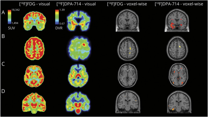
Methods: We compared [18F]FDG and [18F]DPA-714 PET images of 23 patients who had undergone a phase 1 presurgical assessment, using qualitative visual analysis and quantitative analysis, at both the voxel and the regional levels. PET abnormalities (increase in binding for [18F]DPA-714 vs decrease in binding for [18F]FDG) were compared with clinical hypotheses concerning the localization of the EZ based on phase 1 presurgical assessment. The additional value of [18F]DPA-714 PET imaging to [18F]FDG for refining the localization of the EZ was assessed. To strengthen the visual analysis, [18F]DPA-714 PET imaging was also reviewed by 2 experienced clinicians blind to the EZ location.

Results: The study included 23 patients. Visual analysis of [18F]DPA-714 PET was significantly more accurate than [18F]FDG PET to both, show anomalies (95.7% vs 56.5%, p = 0.022), and provide additional information to refine the EZ localization (65.2% vs 17.4%, p = 0.019). All 10 patients with normal [18F]FDG PET had anomalies when using [18F]DPA-714 PET. The additional value of [18F]DPA-714 PET seemed to be greater in patients with normal brain MRI or with neocortical EZ (especially if insula is involved). Regional analysis of [18F]DPA-714 and [18F]FDG PET provided similar results. However, using voxel-wise analysis, [18F]DPA-714 was more effective than [18F]FDG for unveiling clusters whose localization was more often consistent with the EZ hypothesis (87.0% vs 39.1%, p = 0.019). Nonrelevant bindings were seen in 14 of 23 patients in visual analysis and 9 patients of 23 patients in voxel-wise analysis.

Discussion: [18F]DPA-714 PET imaging provides valuable information for presurgical assessments of patients with DRFE. TSPO PET could become an additional tool to help to the localization of the EZ, especially in patients with negative [18F]FDG PET.

Trial registration information: Eudract 2017-003381-27. Inclusion of the first patient: September 24, 2018.

Classification of evidence: This study provides Class IV evidence on the utility of [18F]DPA-714 PET compared with [18F]FDG PET in identifying the epileptic zone in patients undergoing phase 1 presurgical evaluation for intractable epilepsy.


Conflict of interest statement

The authors report no relevant disclosures. Go to Neurology.org/N for full disclosures.

Figures

Figure 1
Figure 1. Additional Value of [18F]DPA-714 PET to Help Localize the EZ
Images shown in anatomical orientation and brain masked, voxel-wise analysis displayed with a p < 0.005, k = 50, uncorrected (brain only), in SPM12, overlaid on a standard MRI section illustrating the decrease of [18F]FDG uptake and increase of [18F]DPA-714 in patient compared with control database. Panel A: The patient with a bitemporal suspected EZ. Visual analysis of [18F]FDG PET shows a bitemporal hypometabolism; the left predominance is not clear. Visual analysis of [18F]DPA-714 PET shows a bitemporal increase of the tracer uptake with a strong left predominance. Voxel-wise analysis of the patient's [18F]FDG PET compared with controls' PET does not identify a statistically significant cluster, whereas [18F]DPA-714 PET voxel-wise analysis identifies a large cluster located in the left temporal gray matter and a smaller one in the right hippocampus. Panel B: The patient with a right pericentral suspected EZ. [18F]DPA-714 PET shows a strong and easily noticeable focal uptake increase within the right precentral area. On retrospective analysis of [18F]FDG PET, a sulcal hypometabolism can be seen within the region pointed by [18F]DPA-714 PET. Voxel-wise analysis of the patient's [18F]FDG and [18F]DPA-714 PET compared with controls' PET identify a statistically significant cluster within the right precentral area. Panel C: The patient with a left insulo-parietal suspected EZ. [18F]FDG PET was initially interpreted as normal. On closer inspection, a slight hypometabolism can be seen within the lesion pointed by [18F]DPA-714 PET. [18F]DPA-714 PET shows an increase in tracer uptake within the left anterior and posterior insula. Voxel-wise analysis of the patient's [18F]FDG PET compared with controls' PET does not identify a statistically significant cluster, whereas [18F]DPA-714 PET voxel-wise analysis identifies a cluster located in the anterior insular gray matter. These images also illustrate the presence of nonrelevant fixation in [18F]DPA-714 PET imaging. Panel D: The patient with a left temporal suspected EZ. Visual analysis of [18F]FDG PET shows a bihippocampal hypometabolism, with a left predominance. Visual analysis of [18F]DPA-714 PET shows an increase of the tracer uptake in both hippocampus with a left predominance and in the left collateral sulcus. Voxel-wise analysis of the patient's [18F]FDG PET compared with controls' PET identifies small cluster in the white matter, whereas [18F]DPA-714 PET voxel-wise analysis identifies a large cluster located in the left collateral sulcus. [18F]DPA-714 = [N,N-diethyl-2-(2-(4-(2[18F]-fluoroethoxy)phenyl)5,7dimethylpyrazolo [1,5a]pyrimidin-3-yl)acetamide; [18F]FDG = [18F]-fluoro-deoxyglucose; EZ = epileptogenic zone; SPM = statistical parametric mapping.
Figure 2
Figure 2. [18F]DPA-714 Uptake Increase, Scored in Qualitative Visual Analysis, Seems to Be Greater With Increased Seizure Frequency (p = 0.049)
[18F]DPA-714 = [N,N-diethyl-2-(2-(4-(2[18F]-fluoroethoxy)phenyl)5,7dimethylpyrazolo [1,5a]pyrimidin-3-yl)acetamide.

References

    1. Kwan P, Schachter SC, Brodie MJ. Drug-resistant epilepsy. N Engl J Med. 2011;365(10):919-926. doi:10.1056/nejmra1004418 - DOI - PubMed
    1. Neuroimaging Subcommision of the International League Against Epilepsy. Commission on diagnostic strategies: recommendations for functional neuroimaging of persons with epilepsy. Epilepsia. 2000;41(10):1350-1356. doi:10.1111/j.1528-1157.2000.tb04617.x - DOI - PubMed
    1. Guidelines for neuroimaging evaluation of patients with uncontrolled epilepsy considered for surgery. Commission on Neuroimaging of the International League Against Epilepsy. Epilepsia. 1998;39(12):1375-1376. doi:10.1111/j.1528-1157.1998.tb01341.x - DOI - PubMed
    1. Wong CH, Bleasel A, Wen L, et al. . Relationship between preoperative hypometabolism and surgical outcome in neocortical epilepsy surgery. Epilepsia. 2012;53(8):1333-1340. doi:10.1111/j.1528-1167.2012.03547.x - DOI - PubMed
    1. Chassoux F, Rodrigo S, Semah F, et al. . FDG-PET improves surgical outcome in negative MRI Taylor-type focal cortical dysplasias. Neurology. 2010;75(24):2168-2175. doi:10.1212/wnl.0b013e31820203a9 - DOI - PubMed

Publication types

Substances

Associated data