. 2023 Nov 7;101(19):e1850-e1862.
doi: 10.1212/WNL.0000000000207794. Epub 2023 Sep 25.

Performance of a [18F]Flortaucipir PET Visual Read Method Across the Alzheimer Disease Continuum and in Dementia With Lewy Bodies

Affiliations

Performance of a [18F]Flortaucipir PET Visual Read Method Across the Alzheimer Disease Continuum and in Dementia With Lewy Bodies

Emma M Coomans et al. Neurology. .

Erratum in

  • Corrections to Received Date Information.
    [No authors listed] [No authors listed] Neurology. 2024 Jul 9;103(1):e209596. doi: 10.1212/WNL.0000000000209596. Epub 2024 Jun 3. Neurology. 2024. PMID: 38830175 Free PMC article. No abstract available.

Abstract

Background and objectives: Recently, the US Food and Drug Administration approved the tau-binding radiotracer [18F]flortaucipir and an accompanying visual read method to support the diagnostic process in cognitively impaired patients assessed for Alzheimer disease (AD). Studies evaluating this visual read method are limited. In this study, we evaluated the performance of the visual read method in participants along the AD continuum and dementia with Lewy bodies (DLB) by determining its reliability, accordance with semiquantitative analyses, and associations with clinically relevant variables.

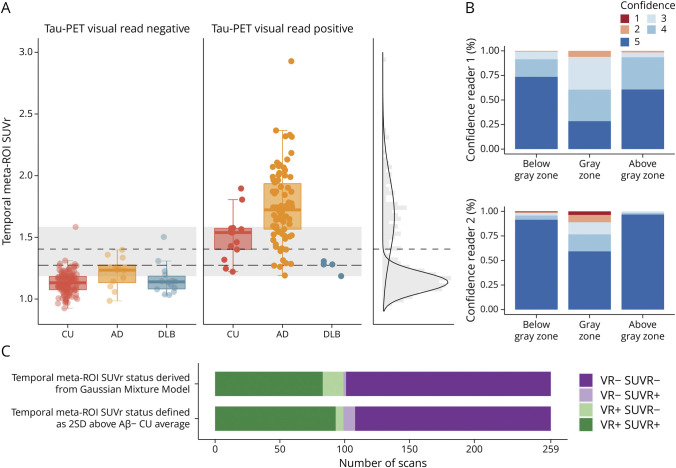
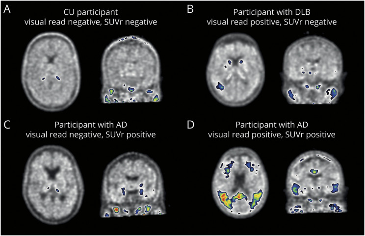
Methods: We included participants who underwent tau-PET at Amsterdam University Medical Center. A subset underwent follow-up tau-PET. Two trained nuclear medicine physicians visually assessed all scans. Inter-reader agreement was calculated using Cohen κ. To examine the concordance of visual read tau positivity with semiquantification, we defined standardized uptake value ratio (SUVr) positivity using different threshold approaches. To evaluate the prognostic value of tau-PET visual read, we performed linear mixed models with longitudinal Mini-Mental State Examination (MMSE).

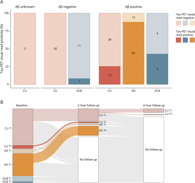
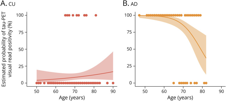
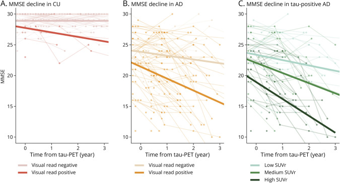
Results: We included 263 participants (mean age 68.5 years, 45.6% female), including 147 cognitively unimpaired (CU) participants, 97 amyloid-positive participants with mild cognitive impairment or AD dementia (AD), and 19 participants with DLB. The visual read inter-reader agreement was excellent (κ = 0.95, CI 0.91-0.99). None of the amyloid-negative CU participants (0/92 [0%]) and 1 amyloid-negative participant with DLB (1/12 [8.3%]) were tau-positive. Among amyloid-positive participants, 13 CU participants (13/52 [25.0%]), 85 with AD (85/97 [87.6%]), and 3 with DLB (3/7 [42.9%]) were tau-positive. Two-year follow-up visual read status was identical to baseline. Tau-PET visual read corresponded strongly to SUVr status, with up to 90.4% concordance. Visual read tau positivity was associated with a decline on the MMSE in CU participants (β = -0.52, CI -0.74 to -0.30, p < 0.001) and participants with AD (β = -0.30, CI -0.58 to -0.02, p = 0.04).

Discussion: The excellent inter-reader agreement, strong correspondence with SUVr, and longitudinal stability indicate that the visual read method is reliable and robust, supporting clinical application. Furthermore, visual read tau positivity was associated with prospective cognitive decline, highlighting its additional prognostic potential. Future studies in unselected cohorts are needed for a better generalizability to the clinical population.

Classification of evidence: This study provides Class II evidence that [18F]flortaucipir visual read accurately distinguishes patients with low tau-tracer binding from those with high tau-tracer binding and is associated with amyloid positivity and cognitive decline.


Conflict of interest statement

E.M. Coomans, L.A. de Koning, R.M. Rikken, S.C.J. Verfaillie, D. Visser, A. den Braber, J. Tomassen, M. van de Beek, and S.S.V. Golla report no competing interests. L.E. Collij has received research support from GE Healthcare (paid to institution). A.W. Lemstra has been funded by ZonMW, Alzheimer Nederland, and Stichting Dioraphte. Research agreements with Combinostics and EPI Pharma. All funding is paid to her institution. A.D. Windhorst is Editor-in-Chief of Nuclear Medicine and Biology. F. Barkhof is in the steering committee or is an iDMC member for Biogen, Merck, Roche, EISAI, and Prothena; consultant for Roche, Biogen, Merck, IXICO, Jansen, and Combinostics. F. Barkhof has research agreements with Merck, Biogen, GE Healthcare, and Roche. F. Barkhof is cofounder and shareholder of Queen Square Analytics LTD. F. Barkhof is supported by the NIHR biomedical research center at UCLH. P.J. Visser is inventor on a patent (#P122938EP10, #P1222938PC00, owner: Stichting VUmc). P. Scheltens has received consultancy fees (paid to the university) from Alzheon, Brainstorm Cell, and Green Valley. Within his university affiliation, he is global PI of the phase 1b study of AC Immune, phase 2b study with FUJI-film/Toyama, and phase 2 study of UCB. He is chair of the EU steering committee of the phase 2b program of Vivoryon, the phase 2b study of Novartis Cardiology, and cochair of the phase 3 study with NOVO-Nordisk. He is also an employee of EQT Life Sciences (formerly LSP). W.M. van der Flier research programs' have been funded by ZonMW, NWO, EU-FP7, EU-JPND, Alzheimer Nederland, Hersenstichting CardioVascular Onderzoek Nederland, Health∼Holland, Topsector Life Sciences & Health, stichting Dioraphte, Gieskes-Strijbis fonds, stichting Equilibrio, Pasman stichting, stichting Alzheimer & Neuropsychiatrie Foundation, Philips, Biogen MA Inc., Novartis-NL, Life-MI, AVID, Roche BV, Fujifilm, and Combinostics. W.M. van der Flier holds the Pasman chair. The chair of W.M. van der Flier is supported by the Pasman stichting. W.M. van der Flier is recipient of ABOARD, which is a public-private partnership receiving funding from ZonMW (#73305095007) and Health∼Holland, Topsector Life Sciences & Health (PPP-allowance; #LSHM20106). W.M. van der Flier has performed contract research for Biogen MA Inc., and Boehringer Ingelheim. W.M. van der Flier has been an invited speaker at Boehringer Ingelheim, Biogen MA Inc., Danone, Eisai, WebMD Neurology (Medscape), and Springer Healthcare. W.M. van der Flier is consultant to Oxford Health Policy Forum CIC, Roche, and Biogen MA Inc. W.M. van der Flier participated in advisory boards of Biogen MA Inc., Roche, and Eli Lilly. All funding is paid to her institution. W.M. van der Flier was associate editor of Alzheimer, Research & Therapy in 2020/2021. W.M. van der Flier is associate editor at Brain. R. Ossenkoppele has given a lecture in a symposium sponsored by GE Healthcare (fee paid to the institution) and is an editorial board member of the European Journal of Nuclear Medicine and Molecular & Imaging of Alzheimer's Research & Therapy. B.N.M. van Berckel has received research support from EU-FP7, CTMM, ZonMw, NWO, and Alzheimer Nederland. B.N.M. van Berckel has performed contract research for Rodin, IONIS, AVID, Eli Lilly, UCB, DIAN-TU, and Janssen. B.N.M. van Berckel was a speaker at a symposium organized by Springer Healthcare. B.N.M. van Berckel has a consultancy agreement with IXICO for the reading of PET scans. B.N.M. van Berckel is a trainer for GE. B.N.M. van Berckel only receives financial compensation from Amsterdam UMC. E. van de Giessen has received research support from NWO, ZonMw, and Hersenstichting. E. van de Giessen has performed contrast research for Heuron Inc., Roche, and 1st Biotherapeutics. E. van de Giessen has a consultancy agreement with IXICO for the reading of PET scans. W.M. van der Flier, R. Ossenkoppele, E. van de Giessen, and A.W. Lemstra are recipients of TAP-Dementia, a ZonMW-funded project (#10510032120003) under the Dutch National Dementia Strategy. Go to Neurology.org/N for full disclosures.

Figures

Figure 1
Figure 1. Cross-sectional and Longitudinal Tau-PET Visual Read Status
(A) The prevalence of baseline tau-PET positivity stratified according to diagnostic group (CU, AD, and DLB) and Aβ status (unknown, negative, and positive) is shown. Numbers indicate the number of participants visually read as tau-negative or tau-positive within each group. (B) Tau-PET visual read status (positive [+] or negative [−]) for each diagnostic group at baseline, 2-year follow-up, and 4-year follow-up indicates that outcome of the visual read method is stable over time. The single CU participant that converted to tau-positive at 4-year follow-up was Aβ positive. Aβ = β-amyloid; AD = Alzheimer disease; CU = cognitively unimpaired; DLB = dementia with Lewy bodies.
Figure 2
Figure 2. Comparing Tau-PET Visual Read With Tau-PET SUVr
(A) Tau-PET SUVr in the temporal meta-ROI is plotted, stratified by diagnostic group (CU, AD, and DLB) and tau-PET visual read status (negative and positive). The short dashed line represents the SUVr cutoff derived from a GMM with the 2 Gaussian distributions plotted on the right. The long dashed line represents the SUVr cutoff defined as 2 SDs above the mean of Aβ negative CU participants. The gray zone represents visual read positive and visual read negative scans with overlapping SUVr. (B) The confidence of the 2 readers (ranging from 1 to 5) is shown for scans below the gray zone, within the gray zone, and above the gray zone. (C) The number of scans with concordant or discordant VR and SUVr status (based both GMM and mean + [2 × SD]) is shown. Aβ = β-amyloid; AD = Alzheimer disease; CU = cognitively unimpaired; DLB = dementia with Lewy bodies; GMM = Gaussian mixture model; ROI = region of interest; SUVr = standardized uptake value ratio; VR = visual read.
Figure 3
Figure 3. Example [18F]Flortaucipir PET Scans for Visual Read
Shown are [18F]flortaucipir PET scans of 4 participants. (A) A CU participant defined as tau-negative on both visual read and SUVr. (B) A participant with DLB defined as visual read tau-positive, but SUVr negative. Increased tracer uptake was observed in only a small region, potentially resulting in low SUVr. (C) A participant with AD defined as visual read negative, but SUVr positive. Increase tracer uptake was observed isolated to the medial temporal lobe, which does not contribute to a positive tau-PET visual read. (D) A participant with AD defined as tau-positive on both visual read and SUVr. AD = Alzheimer disease; CU = cognitively unimpaired; DLB = dementia with Lewy bodies; SUVr = standardized uptake value ratio.
Figure 4
Figure 4. Estimated Probabilities of Tau-PET Visual Read Positivity According to Age
Plotted are the predicted probabilities of tau-PET visual read positivity according to age obtained from a logistic regression between tau-PET visual read (outcome) and age (predictor). We additionally superimposed individual data points to better visualize the distribution of tau-negative and tau-positive cases according to age. A trend toward a positive association between age and probability of tau-PET positivity was observed in CU participants (A), whereas a strong negative association between age and probability of tau-PET positivity was observed in participants with AD (B). AD = Alzheimer disease; CU = cognitively unimpaired.
Figure 5
Figure 5. Association of Tau-PET Visual Read and Tau-PET SUVr With Prospective MMSE
Spaghetti plots of longitudinal MMSE are shown. Association of tau-PET visual read status with longitudinal performance on the MMSE is shown for (A) CU participants and (B) participants with AD, with slopes from linear mixed models superimposed on the graphs. In (C) the association of temporal meta-ROI, tau-PET SUVr with longitudinal performance on the MMSE in visual read tau-positive participants with AD is shown. For visualization purposes, slopes from linear mixed models with SUVr in tertiles are superimposed on the graph. AD = Alzheimer disease; CU = cognitively unimpaired; MMSE = Mini-Mental State Examination; ROI = region of interest; SUVr = standardized uptake value ratio.

Comment in

References

    1. Scheltens P, De Strooper B, Kivipelto M, et al. . Alzheimer's disease. Lancet. 2021;397(10284):1577-1590. doi:10.1016/S0140-6736(20)32205-4 - DOI - PMC - PubMed
    1. de Wilde A, van der Flier WM, Pelkmans W, et al. . Association of amyloid positron emission tomography with changes in diagnosis and patient treatment in an unselected memory clinic cohort: the ABIDE project. JAMA Neurol. 2018;75(9):1062-1070. doi:10.1001/jamaneurol.2018.1346 - DOI - PMC - PubMed
    1. Duits FH, Prins ND, Lemstra AW, et al. . Diagnostic impact of CSF biomarkers for Alzheimer's disease in a tertiary memory clinic. Alzheimers Dement. 2015;11(5):523-532. doi:10.1016/j.jalz.2014.05.1753 - DOI - PubMed
    1. Ossenkoppele R, Rabinovici GD, Smith R, et al. . Discriminative accuracy of [18F]flortaucipir positron emission tomography for Alzheimer disease vs other neurodegenerative disorders. JAMA. 2018;320(11):1151-1162. doi:10.1001/jama.2018.12917 - DOI - PMC - PubMed
    1. Jack CR, Wiste HJ, Botha H, et al. . The bivariate distribution of amyloid-beta and tau: relationship with established neurocognitive clinical syndromes. Brain. 2019;142(10):3230-3242. doi:10.1093/brain/awz268 - DOI - PMC - PubMed

Publication types