

Case Reports
doi: 10.1016/j.abd.2022.06.008.
Epub 2023 Sep 23.
Pigmented squamous cell carcinoma in a non-photo-exposed area of an indigenous woman
Affiliations
- PMID: 37749019
- PMCID: PMC10964365
- DOI: 10.1016/j.abd.2022.06.008
Item in Clipboard
Case Reports
Pigmented squamous cell carcinoma in a non-photo-exposed area of an indigenous woman
An Bras Dermatol.
2024 Jan-Feb.
No abstract available
Figures
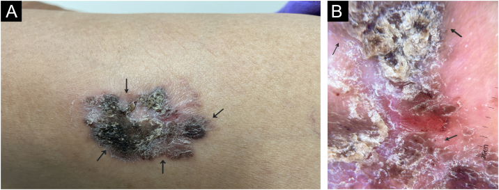
(A) On examination, an infiltrated erythematous-brownish plaque was observed, surmounted by a hyperkeratotic area, in the proximal lateral region of the left thigh (covered area). (B) Dermoscopy showing erythema and linear vessels in the central region, areas of black pigment and glomerular vessels and radiated pigment in the periphery.
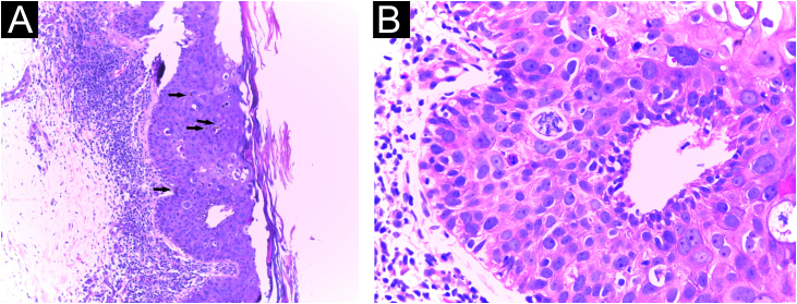
Histopathology of an incisional biopsy, which suggested the diagnosis of pigmented SCC in situ. (A) Compact hyperkeratosis, acanthosis and pigment deposits, in addition to atypical keratinocytes and mitoses in the middle portion of the epidermis (Hematoxylin & eosin, ×40). (B) At higher magnification, atypical keratinocytes and mitoses (Hematoxylin & eosin, ×400).
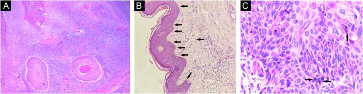
Histopathology of the excisional biopsy. (A) Invasive cutaneous neoplasm consisting of atypical and pleomorphic cells with clear eosinophilic cytoplasm and pleomorphic nuclei with abundant keratinization (Hematoxylin & eosin, ×100). (B) Pigment deposits (Hematoxylin & eosin, ×40). (C) At higher magnification, keratinocyte atypia and pigment deposits (Hematoxylin & eosin, ×400).

Immunohistochemistry. (A) Positivity for EMA (epithelial membrane antigen). (B) Positivity for p63 protein.
References
-
- gbm.org [Internet]. Cartilha de tratamento – CEC de pele. 2019. Grupo Brasileiro de Melanoma. [Cited 2022 Mar 4]. Available from: https://gbm.org.br/wp-content/uploads/2019/09/livreto-GBM-v2.pdf.
-
- Satter E.K. Pigmented squamous cell carcinoma. Am J Dermatopathol. 2007;29:486–489. - PubMed
-
- Jeunon T., Vita-Campos C.M., Azeredo-Coutinho R.B. Case for diagnosis: pigmented squamous cell carcinoma. An Bras Dermatol. 2009;84:293–295. - PubMed
-
- Dimitra K., Vassiliki Z., Maria G., Chrisoula S. Pigmented squamous cell carcinoma of the lower back skin: a case report and review of the literature. Pigmentary Disorders. 2015;2:165.
Publication types
MeSH terms
LinkOut - more resources
Full Text Sources
Medical
