. 2023 Sep 25;14(1):5964.
doi: 10.1038/s41467-023-41734-4.

An original potentiating mechanism revealed by the cryo-EM structures of the human α7 nicotinic receptor in complex with nanobodies

Affiliations

An original potentiating mechanism revealed by the cryo-EM structures of the human α7 nicotinic receptor in complex with nanobodies

Marie S Prevost et al. Nat Commun. .

Abstract

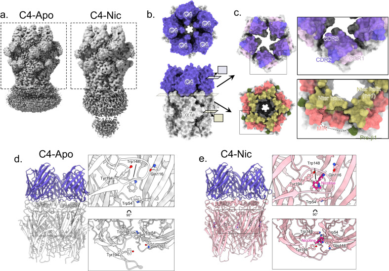
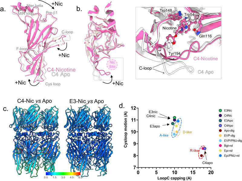
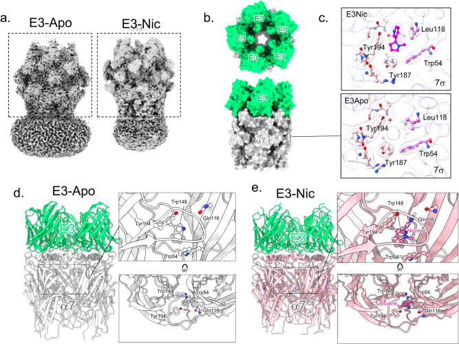
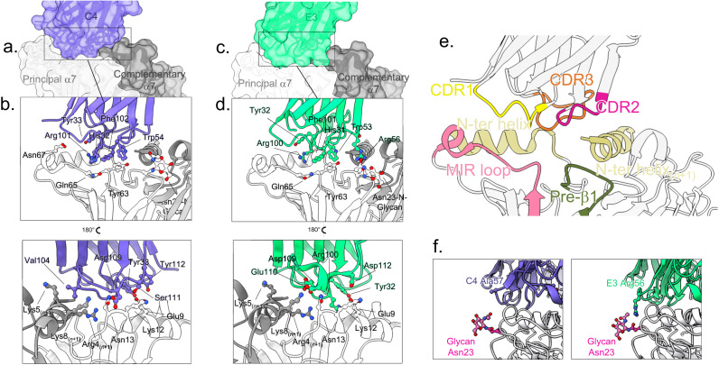
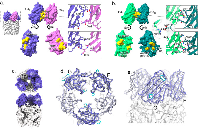
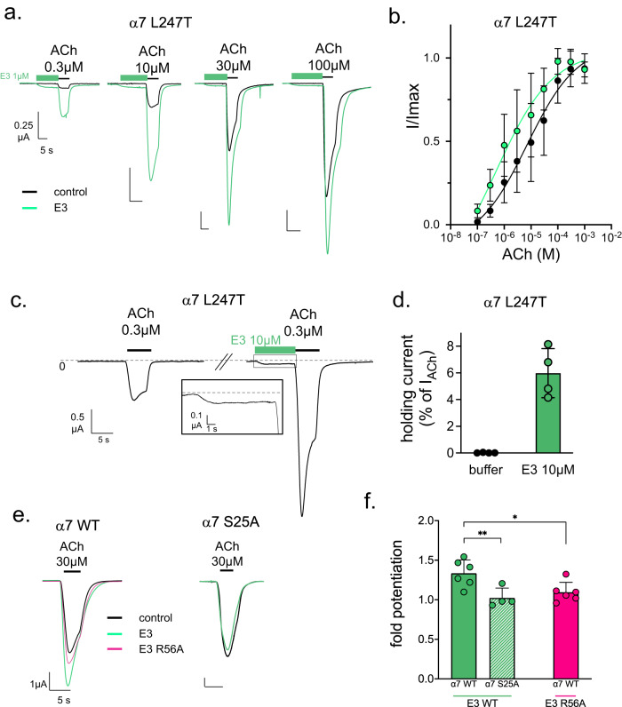
The human α7 nicotinic receptor is a pentameric channel mediating cellular and neuronal communication. It has attracted considerable interest in designing ligands for the treatment of neurological and psychiatric disorders. To develop a novel class of α7 ligands, we recently generated two nanobodies named E3 and C4, acting as positive allosteric modulator and silent allosteric ligand, respectively. Here, we solved the cryo-electron microscopy structures of the nanobody-receptor complexes. E3 and C4 bind to a common epitope involving two subunits at the apex of the receptor. They form by themselves a symmetric pentameric assembly that extends the extracellular domain. Unlike C4, the binding of E3 drives an agonist-bound conformation of the extracellular domain in the absence of an orthosteric agonist, and mutational analysis shows a key contribution of an N-linked sugar moiety in mediating E3 potentiation. The nanobody E3, by remotely controlling the global allosteric conformation of the receptor, implements an original mechanism of regulation that opens new avenues for drug design.


Conflict of interest statement

G.A., P.J.C., P.L., M.S.P. and N.B. are inventors of patent application US 63/383,099 that covers the nanobodies and therapeutic uses thereof. The remaining authors declare no competing interests.

Figures

Fig. 1
Fig. 1. C4-α7 cryo-EM structures in the absence or presence of nicotine.
a Unsharpened maps of the α7∆ICDcryo in complex with C4 (C4-Apo, left) and of the α7FLcryo in complex with C4 and nicotine (C4-Nic, right) with densities contoured at 2σ. The dashed boxes denote the parts that were used for model building. b Top and side views of the C4-Apo structure, protein chains are depicted in white (α7 apo-) and purple (C4). The structure is shown exploded in (c). c Top: Bottom view of the ring of C4 with the molecular surface of the three CDRs colored in shades of purple. Bottom: Top view of the ECD of α7 with N-ter helices, MIR and pre-β1 colored in yellow, pink and green, respectively. The groove in which the CDRs are plunging is delimited with a dashed line. d Side view of the C4-Apo structure, represented in cartoon with α7 in white and C4 in purple. The orthosteric binding site is enlarged and further rotated by 90 °C in the right panels. Residues involved in nicotine binding are depicted in sticks. e Side view of the C4-Nic structure, represented in cartoon with α7 in pink and C4 in purple. The orthosteric binding site is enlarged and further rotated by 90 °C in the right panels. Residues involved in nicotine binding and nicotine itself are depicted in sticks.
Fig. 2
Fig. 2. α7 conformation in C4-Apo corresponds to an apo-like ECD conformation, and in C4-Nic, E3-Apo and E3-Nic to an agonist-bound ECD conformation.
a α7 monomers from the C4-apo (gray) and C4-Nic structure (pink) are seen from the side and depicted in cartoons after superimposition of the whole α7 pentamer. Landmark loops are labeled, and motions from Apo- to Nic- are highlighted with black arrows. b The same representation as in (a) but seen from the top. The nicotine binding site is depicted with a pink circle. A close view of the nicotine binding site is shown on the right. Nicotine and key residues from the binding pocket are labeled and depicted in sticks. c Conformational changes upon nicotine binding in C4-bound (left) and E3-bound (right) structures. C4-Nic and E3-Nic are represented in cartoons, and Cα are colored according to the rmsd value (in Å) calculated using their Apo- counterpart, with the same range for both and shown in the color key below. d Correlation of the cys-loop outward motion (in Å) and Loop C capping (in Å) in the known structures of α7 allows to define pairs of values that are landmarks of the α7 conformations. R, A, and D-like structures are grouped in dashed circles.
Fig. 3
Fig. 3. E3-α7 cryo-EM structures in the absence or presence of nicotine.
a Unsharpened maps of the α7∆ICDcryo in complex with E3-Apo (left) and E3-Nic (right) with densities contoured at 2σ. The dashed boxes denote the parts that were used for model building. b Top and side views of the E3-Apo structure, protein chains are depicted in white (α7 apo-) and green (E3). The orthosteric sites are shown in (c). c Neurotransmitter binding pocket of E3-Nic and E3-Apo. Electron densities are contoured at 7σ, and residues involved in neurotransmitter binding are shown in sticks. Nicotine was built in the extra density found in E3Nic, while the small and spherical density found in E3-Apo likely fits a cation or a water molecule. d Side view of the E3-Apo structure, represented in cartoon with α7 in white and E3 in green. The orthosteric binding site is enlarged and further rotated by 90 °C in the right panels. Residues involved in nicotine binding are depicted in sticks. e Side view of the E3-Nic structure, represented in cartoon with α7 in pink and E3 in green. The orthosteric binding site is enlarged and further rotated by 90 °C in the right panels. Residues involved in nicotine binding and nicotine itself are depicted in sticks.
Fig. 4
Fig. 4. Binding pose and epitope of C4 and E3.
a Overview of the binding site of C4 on α7 exemplified by the C4-Apo structure. One C4 molecule (purple surface and cartoon) contacts two α7 subunits depicted in light (principal n subunit) and dark (complementary n+1 subunit) surface and cartoons. b Details of the binding site as seen from the solvent (upper panel) or the vestibule (lower panel) of α7 with color code as in (a). key residues are labeled, and their side chains displayed in stick balls. c Same as in (a) but with the E3-Apo structure, E3 is colored in green. d Same as in (b). but with the E3-Apo structure, E3 is colored in green. e Overview of the loops involved in binding exemplified by the C4-Apo structure represented in cartoons. f Interaction of E3 with Asn23 glycan. Close view of the Glycan (pink sticks) built on α7-Asn23 (pink sticks) from the pre-β1 of α7 (gray) and of the CDR2 of C4 (left, purple) or E3 (right, green). Ala57 from C4 and its homologous Arg56 from E3 are depicted in sticks.
Fig. 5
Fig. 5. Nanobody-nanobody interactions.
a Surface representation of two adjacent C4 (F in purple, G in pink) in C4-Apo and their exploded view below. Residues at the interface are colored in yellow, and their details are shown in cartoons and sticks in a close-up view (right). b Surface representation of two adjacent E3 (F in light green, G in dark pink) in E3-Apo and their exploded view below. Residues at the interface are colored in yellow, and their details are shown in cartoons and sticks in a close-up view (right). Additionally, the putative salt bond between Arg108 and Asp113 is detailed in a separate close-up view, with the density contoured at 4σ. c Overview of the C4partial-Apo structure from the top (top) and the side (bottom) with α7 in gray and the three C4 molecules in purple. d Superimposition of the C4partial-Apo on the C4-Apo structures, aligned on α7 ECD. Both structures are represented in ribbon, α7 in gray, C4 from C4partial-Apo in purple and C4 from C4-Apo in gray blue. Motion from C4-Apo to C4partial-Apo are highlighted with arrows. On the left, structures are seen from the top and show a lateral inclination of the nanobodies toward the anticlockwise subunit accompanied by a radial inclination toward the vestibule. e Same as in (d), but with structures seen from the side, with the most displaced nanobody in front (chain G).
Fig. 6
Fig. 6. Electrophysiological analysis of the potentiation by E3.
a Representative traces of ACh dose-response curves of the α7 L247T mutant with (green) or without (black) 15-s pre-application of 1 µM E3 on a single cell. b Resulting dose-response curves after normalization on maximal currents of both conditions. Points are mean ± s.d. with n = 11 cells. c E3 direct activation on α7 L247T mutant recorded by TEVC on Xenopus oocytes. Representative trace showing a first 0.3 µM ACh application yielding slow-desensitizing currents. After 2-min washing, 10 µM E3 was applied for 10 s, evoking significant current, before 0.3 µM ACh. A close-up view of the recording during E3 application is shown in a black box. d Holding currents measured on α7 L247T with or without 10 µM E3 normalized with the non-potentiated response to 0.3 µM ACh. Points are mean ± s.d. with n = 4 cells. e Potentiation assays of E3-WT and E3-R56A on α7 WT and of E3-WT on α7-S25A. Responses to 30 µM ACh in the absence of E3 (black) are superimposed with response after 30 s application of 1 µM of E3 wild-type (green) or R56A (pink). f Fold potentiation calculated on n = 4 to 6 cells by E3-WT on α7 WT and S25A mutant and of E3-R56A on α7-WT. Bars are mean ± s.d. Values were submitted to an unpaired t-test with *p = 0.0183, **p = 0.0013.

Similar articles

Cited by

References

    1. Nemecz A, Prevost MS, Menny A, Corringer P-J. Emerging molecular mechanisms of signal transduction in pentameric ligand-gated ion channels. Neuron. 2016;90:452–470. doi: 10.1016/j.neuron.2016.03.032. - DOI - PubMed
    1. Papke RL, Horenstein NA. Therapeutic targeting of α 7 nicotinic acetylcholine receptors. Pharmacol. Rev. 2021;73:1118–1149. doi: 10.1124/pharmrev.120.000097. - DOI - PMC - PubMed
    1. Hone AJ, McIntosh JM. Nicotinic acetylcholine receptors in neuropathic and inflammatory pain. FEBS Lett. 2018;592:1045–1062. doi: 10.1002/1873-3468.12884. - DOI - PMC - PubMed
    1. Terry, A. V. α7 nicotinic acetylcholine receptors as therapeutic targets in schizophrenia: update on animal and clinical studies and strategies for the future. Neuropharmacology170, 108053 (2020). - PMC - PubMed
    1. Young GT, Zwart R, Walker AS, Sher E, Millar NS. Potentiation of α7 nicotinic acetylcholine receptors via an allosteric transmembrane site. Proc. Natl Acad. Sci. USA. 2008;105:14686–14691. doi: 10.1073/pnas.0804372105. - DOI - PMC - PubMed

Publication types

Substances