. 2023 Sep 25;13(1):15992.
doi: 10.1038/s41598-023-42797-5.

Immunomodulatory effects of nanoparticles on dendritic cells in a model of allergic contact dermatitis: importance of PD-L2 expression

Affiliations

Immunomodulatory effects of nanoparticles on dendritic cells in a model of allergic contact dermatitis: importance of PD-L2 expression

Angela Wong Lau et al. Sci Rep. .

Abstract

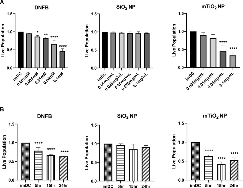
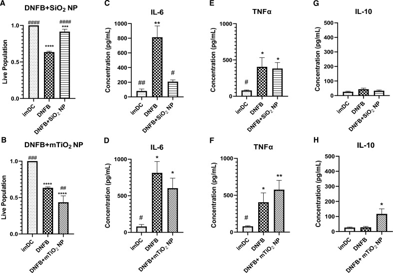
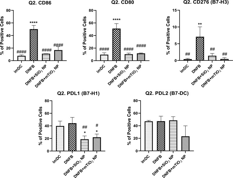
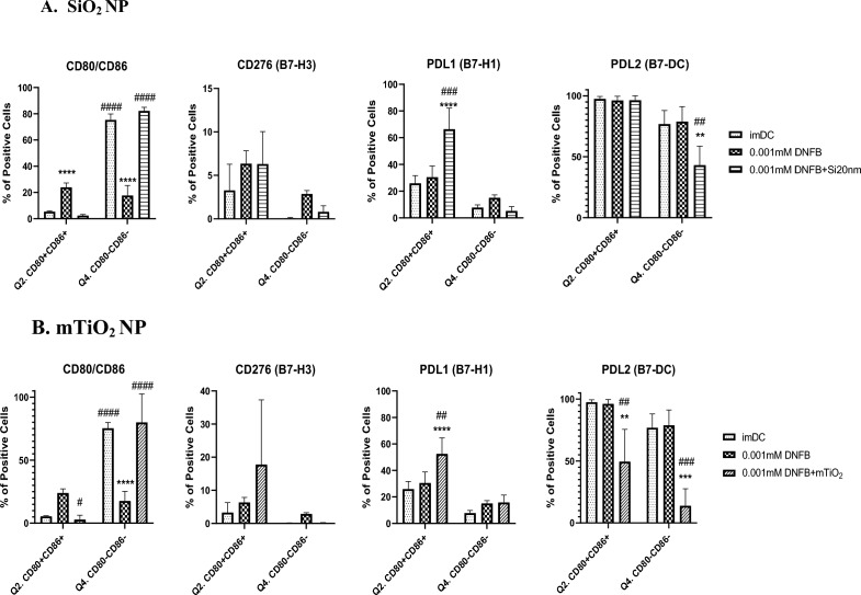
Nanoparticle (NP) skin exposure is linked to an increased prevalence of allergic contact dermatitis. In our prior studies using the mouse contact hypersensitivity (CHS) model, we reported that silica 20 nm (SiO2) NPs suppressed the allergic response and titanium dioxide NPs doped with manganese (mTiO2) exacerbated it. In this work, we conducted in vitro experiments using bone marrow-derived dendritic cells (BMDCs) to study the combinatorial effect of the potent 2,4-dinitrofluorobenzene (DNFB) hapten sensitizer with SiO2 and mTiO2 NPs on BMDC cytotoxicity, cytokine secretion and phenotype using the B7 family ligands. Results show that DNFB and mTiO2 behave similarly and exhibit proinflammatory characteristics while SiO2 promotes a naive phenotype. We observe that the B7-H3 (CD276) ligand is only expressed on CD80 + (B7-1) BMDCs. Results from adoptive transfer CHS studies, combined with BMDC phenotype analysis, point to the importance of PD-L2 expression in modulating the adaptive immune response. This work identifies metrics that can be used to predict the effects of NPs on contact allergy and to guide efforts to engineer cell-based therapies to induce hapten specific immune tolerance.


Conflict of interest statement

The authors declare no competing interests.

Figures

Figure 1
Figure 1
Effects of DNFB, SiO2 and mTiO2 exposure on BMDC cytotoxicity as a function of concentration and time. BMDCs were harvested on day 8 and treated with DNFB, SiO2 and mTiO2 to study cytotoxicity as a function of concentration and time by flow cytometry. (A) Cell viability as a function of concentration for a 1 h exposure. (B) Cell viability following exposure to the lowest non-cytotoxic concentrations from (A) and exposed over a period of 5 h, 15 h and 24 h. Results indicate that cytotoxicity of DNFB and mTiO2 NPs on BMDCs was dose- and time- dependent. Live population was normalized the imDC no treatment control. Ordinary one-way ANOVA was performed and compared to imDC. N = 3–5. Mean ± SD. *p < 0.05, **p < 0.01, ***p < 0.001, ****p < 0.0001.
Figure 2
Figure 2
Effects of DNFB, SiO2 and mTiO2 exposure on BMDC cytokine secretion. BMDCs were exposed to a low non-cytotoxic concentrations of each stressor (0.001 mM DNFB, 0.01 mg/mL SiO2 and 0.005 mg/mL mTiO2) over a period of 5 h, 15 h and 24 h. ELISA was used to analyze cytokines in cell culture supernatants: (A) IL-6, (B) TNFα, (C) IL-10. All stressors tend to increase IL-6 and TNFα. Only mTiO2 increased IL-10. Concentration was normalized against % of live cells. Ordinary one-way ANOVA was performed and compared to imDC. N = 3–5. Mean ± SD. *p < 0.05, **p < 0.01, ***p < 0.001, ****p < 0.0001.
Figure 3
Figure 3
Co-stimulatory molecules from the B7 family quantified by flow cytometry gated on the CD11c + MHCII + subpopulation following BMDC exposure to each stressor over time. The phenotypic characteristics of BMDCs following exposure to a low concentration of each stressor (0.001 mM DNFB, 0.01 mg/mL SiO2 and 0.005 mg/mL mTiO2) was followed over time by flow cytometry gated under the CD11c+ MHCII + (Q2.) subpopulation. DNFB and mTiO2 behave more similar with upregulation of CD86, CD276, and PD-L1 over time. Only DNFB upregulated CD80. Exposure to SiO2 did not alter these B7 ligands over time. Ordinary one-way ANOVA was performed and compared to imDC. N = 3–5. Mean ± SD. *p < 0.05, **p < 0.01, ***p < 0.001, ****p < 0.0001.
Figure 4
Figure 4
Co-stimulatory molecules from the B7 family quantified by flow cytometry gated on the CD11c+ MHCII+ and CD80/CD86 subpopulations following BMDC exposure to each stressor over time. Results for DNFB are colored in blue, SiO2 in green and mTiO2 in purple. The phenotypic characteristics of BMDCs following exposure to a low concentration of each stressor (0.001 mM DNFB, 0.01 mg/mL SiO2 and 0.005 mg/mL mTiO2) was followed over time, 5 h, 15 h, and 24 h, by flow cytometry gated under the CD11c + MHCII + CD80/CD86 subpopulations. Presented here are changes the CD80 + CD86+ double positive (DP) (Q2.) and the CD80-CD86- double negative (DN) (Q4.) subpopulations including the expression levels of PD-L1 (B7-H1), PD-L2 (B7-DC) and CD276 (B7-H3) on the DP and DN subpopulations. For DNFB exposure, the activated DP population clearly increases as the naive DN decreases and PD-L1 expression increased on the DN at 24 h. mTiO2 exposure over time parallels the effects of DNFB exposure. In contrast, SiO2 exposure over time tended to decrease the DP subpopulation and it significantly decreased PD-L1 expression in the DP subpopulation. Both SiO2 and mTiO2 tended to increased CD276 in the DP at 24 h. The similarities between DNFB and mTiO2 were evident in CD80/CD86 and PD-L1 expression and distinct from SiO2. A two-way ANOVA was performed and compared to imDC. N = 3–5. Mean ± SD. *p < 0.05, **p < 0.01, ***p < 0.001, ****p < 0.0001.
Figure 5
Figure 5
BMDC cytotoxicity and cytokine secretion following co-exposure to DNFB with SiO2 or mTiO2 NPs. BMDCs were co-exposed to DNFB plus SiO2 or mTiO2 nanoparticles for 24 h. Cell viability and secreted cytokines were measured and compared to DNFB treatment alone and to untreated imDC as a control. (A) SiO2 NPs were shown to have cytoprotective effect in DNFB co-exposure whereas (B) mTiO2 NPs induced higher cytotoxicity. Supernatants were collected to analyze cytokine production by ELISA for (C,D) IL-6 which showed co-culture with SiO2 NPs decreased IL-6 production while co-culture with mTiO2 did not. (E,F) addition of either NPs did not alter the levels of TNFα produced by DNFB alone and (G,H) the co-culture of DNFB with mTiO2 NPs increased IL-10 production while SiO2 NPs did not. Ordinary one-way ANOVA was performed and compared to imDC (*) and DNFB alone (#). Concentration was normalized against % of live cells. N = 3–5. Mean ± SD. */#p < 0.05, **/##p < 0.01, ***/###p < 0.001, ****/####p < 0.0001.
Figure 6
Figure 6
Co-stimulatory molecules from the B7 family quantified by flow cytometry gated on the CD11c + MHCII + subpopulation following co-exposure of DNFB with NPs. BMDCs were treated with DNFB (0.001 mM DNFB) and co-exposed with SiO2 (0.01 mg/mL) or mTiO2 (0.005 mg/mL) NPs for 24 h. Surprisingly, both NPs appeared to suppress the activation of all co-stimulatory molecules relative to DNFB only at 24 h, except for PD-L2, for which mTiO2 significantly down regulated it. Ordinary one-way ANOVA was performed and compared to imDC (*) and DNFB (#). N = 3–5. Mean ± SD.*/#p < 0.05, **/##p < 0.01, ***/###p < 0.001, ****/####p < 0.0001.
Figure 7
Figure 7
Co-stimulatory molecules from the B7 family quantified by flow cytometry gated on the CD11c + MHCII + CD80/CD86 subpopulations following 24 h DNFB alone or DNFB plus NP co-exposure. CD80/CD86 subpopulations were divided and CD276 (B7-H3), PD-L1 (B7-H1) and PD-L2 (B7-DC) were gated under, double positive (DP) population (Q2.) or double negative (DN) population (Q4.) to determine whether their expression varies under the different activation states of BMDCs by using the activation markers CD80/CD86 following non-cytotoxic concentrations of (A) 0.001 mM DNFB alone and plus 0.01 mg/mL SiO2 co-exposure and (B) 0.001 mM DNFB alone and pls 0.005 mg/mL mTiO2 co-exposure. NPs suppressed BMDCs activation, increased PD-L1 expression and mTiO2 induced a significant decrease in PD-L2 expression in the DP subpopulation at 24 h while SiO2 did not. A two-way ANOVA was performed and compared to imDC (*) and DNFB (#). N = 3–5. Mean ± SD. **/##p < 0.01, ***/###p < 0.001, ****/####p < 0.0001.
Figure 8
Figure 8
Comparison of CHS allergic response for different models of sensitization and challenge. (A) Comparison of ear swelling response for DNFB topical (0.05%) vs. S.C. (0.01 mM DNFB) sensitization with 0.2% DNFB challenge on one ear vs. vehicle on the other. No differences between these sensitization methods was observed. (B) Comparison of ear swelling response for S.C. sensitization with DNFB only and challenge with DNFB or DNFB co-exposure with NPs and vehicle on the other. Ear swelling was exacerbated with mTiO2 NPs but decreased with SiO2 NPs compared to DNFB alone. Ordinary one-way ANOVA was performed and compared to imDC (*). N = 3–12. Mean ± SD. *p < 0.05, **p < 0.01. (C) Comparison of ear swelling response for S.C. sensitization with BMDC treated 1 h with DNFB only or DNFB + SiO2 or DNFB + mTiO2 and challenge with 0.2% DNFB on one ear vs. vehicle on the other. Ear swelling was exacerbated with mTiO2 but decreased with SiO2 compared to DNFB alone.

Update of

Similar articles

Cited by

References

    1. Salah S, Taieb C, Demessant AL, Haftek M. Prevalence of skin reactions and self-reported allergies in 5 countries with their social impact measured through quality of life impairment. Int. J. Environ. Res. Public Health. 2021 doi: 10.3390/ijerph18094501. - DOI - PMC - PubMed
    1. Owen JL, Vakharia PP, Silverberg JI. The role and diagnosis of allergic contact dermatitis in patients with atopic dermatitis. Am. J. Clin. Dermatol. 2018;19:293–302. doi: 10.1007/s40257-017-0340-7. - DOI - PMC - PubMed
    1. Thyssen JP, Linneberg A, Menne T, Johansen JD. The epidemiology of contact allergy in the general population–prevalence and main findings. Contact Dermat. 2007;57:287–299. doi: 10.1111/j.1600-0536.2007.01220.x. - DOI - PubMed
    1. Nosbaum A, Vocanson M, Rozieres A, Hennino A, Nicolas JF. Allergic and irritant contact dermatitis. Eur. J. Dermatol. 2009;19:325–332. doi: 10.1684/ejd.2009.0686. - DOI - PubMed
    1. Roberts DW. Allergic contact dermatitis: Is the reactive chemistry of skin sensitizers the whole story? A response. Contact Dermat. 2013;68:245–249. doi: 10.1111/cod.12057. - DOI - PubMed

Publication types