. 2023 Sep 25;13(1):16057.
doi: 10.1038/s41598-023-42997-z.

Discovery of Nedd4 auto-ubiquitination inhibitors

Affiliations

Discovery of Nedd4 auto-ubiquitination inhibitors

Darren Yong et al. Sci Rep. .

Abstract

E3 ubiquitin ligases are critical to the protein degradation pathway by catalyzing the final step in protein ubiquitination by mediating ubiquitin transfer from E2 enzymes to target proteins. Nedd4 is a HECT domain-containing E3 ubiquitin ligase with a wide range of protein targets, the dysregulation of which has been implicated in myriad pathologies, including cancer and Parkinson's disease. Towards the discovery of compounds disrupting the auto-ubiquitination activity of Nedd4, we developed and optimized a TR-FRET assay for high-throughput screening. Through selective screening of a library of potentially covalent compounds, compounds 25 and 81 demonstrated apparent IC50 values of 52 µM and 31 µM, respectively. Tandem mass spectrometry (MS/MS) analysis confirmed that 25 and 81 were covalently bound to Nedd4 cysteine residues (Cys182 and Cys867). In addition, 81 also adducted to Cys627. Auto-ubiquitination assays of Nedd4 mutants featuring alanine substitutions for each of these cysteines suggested that the mode of inhibition of these compounds occurs through blocking the catalytic Cys867. The discovery of these inhibitors could enable the development of therapeutics for various diseases caused by Nedd4 E3 ligase dysregulation.


Conflict of interest statement

The authors declare no competing interests.

Figures
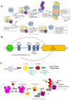
Figure 1
Figure 1
Schematics of Nedd4 function, domain structure and a TR-FRET assay for monitoring auto-ubiquitination. (A) The role of Nedd4 E3 ligase in degradation of target proteins and modulation of its activity by small molecule inhibitors is depicted. (B) Nedd4 has 3 types of folded domains. The C2 domain (20–122) drawn as a green circle, the WW domains; WW1 (192–224), WW2 (349–381), WW3 (422–454) and WW4 (474–506) drawn as blue boxes and the HECT domain (563–899) drawn as orange rectangle. The positions of the domains were predicted using the Simple Modular Architecture Research Tool (SMART) online resource by querying the full-length (1–900) Nedd4 isoform 1 sequence (NCBI accession: NP_006145.2). The sequence of the inhibitory linker peptide (225–244) between WW1 and WW2 is shown below. The red lines indicate intramolecular protein interactions with the HECT domain that inhibit E3 ligase activity. (C) When the terbium (Tb) labelled SA becomes excited by light at 340 nm (represented by purple in the diagram), Tb emits light at 480 nm (blue). The 480 nm emission serves as the excitation source for the FITC labelled Ub and emits light at 528 nm (green). Upon E3 ubiquitin conjugation, the terbium and FITC are brought in close proximity (within 10 nm) to allow for FRET pairing.
Figure 2
Figure 2
Assessment of TR-FRET signal for detecting Nedd4 auto-ubiquitination activity. (A) Plotting TR-FRET emission ratio above background (528/485 nm) of the full-length WT (1–900) and truncated variants (153–900) lacking the inhibitory C2 domain with and without the addition of 2 mM ATP-MgCl2. The mutant variant of the truncated protein also lacks the inhibitory linker peptide (225–244). (B) Removing reaction components demonstrates a consistent decrease in the TR-FRET 528/485 nm emission ratio with the loss of one or more reagents, demonstrating that all reaction components are essential for ubiquitination to occur during the assay. All values are from the experiments performed in triplicate, and are presented as mean ± standard deviation. The composition of each reaction (1 to 11) is presented in the corresponding column in the table below the numbers.
Figure 3
Figure 3
Initial screening of compounds for full-length Nedd4 auto-ubiquitination inhibition. (A) The first set of compounds were screened at 500 µM. (B) The second set of compounds were screened at 300 µM. (C) The final set of compounds were screened at 200 µM. Screening of compounds were performed in triplicate, and are presented as mean ± standard deviation. Information on each set of compounds is presented in supplementary Table 3 and Source Data 1. Percent activity was calculated as ratio of the background subtracted TR-FRET signal at any given concentration of a potential inhibitor and the background subtracted TR-FRET signal in the absence of compound.
Figure 4
Figure 4
Covalent inhibitors of full-length Nedd4 auto-ubiquitination. Compounds were chosen from the initial screen when demonstrating > 50% inhibition at the single high concentration initially tested. Experiments were performed in triplicate following a 2 h RT incubation in the selected compound at varying concentrations with data presented as the relative auto-ubiquitination activity compared to the uninhibited conditions. Data were analyzed and plotted using 4-parameter non-linear regression. The relative IC50 values ± standard deviation are displayed on curves whose data followed a clear sigmoidal response over the tested concentration range. Catalog numbers for the compounds (and arbitrary number in parentheses) are shown below the graph and molecular structures are presented alongside the data. All compounds were obtained from Enamine with the exception of 3 (QD-1755) which was purchased from Combi-Blocks. Information on all compounds is presented in supplementary Table 3 and Source Data 1.
Figure 5
Figure 5
Effects of compound preincubation on the potency of inhibitors. Activities measured by difference in emission ratio at 528/485 nm between reactions with and without enzyme, are presented relative to the uninhibited assay in 1% DMSO (100% activity). Assays were run with or without a 2 h preincubation in 200 µM of inhibitor prior to initiation of the auto-ubiquitination reaction. Auto-ubiquitination reactions were allowed to proceed for 1 h at 37 °C after the addition of 2 mM ATP-MgCl2. Data represent means of three reactions performed in separate microplate wells ± standard deviation. Data for each compound were analyzed and compared to the relative activity without the preincubation through a one-way ANOVA with Dunnett's post hoc test applied. When the inhibition associated with one of the 2 h incubation conditions is significantly different (p < 0.05) from the control without preincubation the data is labeled with a '*'.
Figure 6
Figure 6
Confirmation of the inhibitory effect of compound 25 and 81 on Nedd4 auto-ubiquitination by Western Blot. Nedd4 auto-ubiquitination was monitored by (A) using fluorescently labeled streptavidin to visualize biotinylated full-length Nedd4, and (B) mouse anti-ubiquitin antibody and fluorescently labeled donkey anti-mouse secondary IgG antibody combination to detect ubiquitin. Anti-ubiquitin signal is shown in red and labeled streptavidin in blue. (C) The overlay of A and B is shown. Inhibition by two inhibitors 81 and 25 demonstrates differing potencies with 81 showing a complete loss of ubiquitination on the Nedd4 band at the two highest concentrations tested. However, 25 shows a modest loss of activity at 200 μM, most clearly indicated by the increase in the levels of free ubiquitin following the reaction in B and C. The samples in wells labeled 1 to 9 are as follows: (1) Protein molecular weight standard, (2) Reaction components minus ATP-MgCl2 (negative control), (3) Uninhibited reaction (positive control), (4) 10 µM 81, (5) 50 µM 81, (6) 100 µM 81, (7) 50 µM 25, (8) 100 µM 25, (9) 200 µM 25.
Figure 7
Figure 7
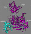
Visualization of the cysteine site locations in the Nedd4 HECT domain that are confirmed targets for adduction by compounds 25 and 81 in relation to the Ub exosite. Structure visualization of Nedd4-1 was performed using ChimeraX software on a solved crystal structure of human Nedd4 HECT domain bound to Ub in the Exo-Ub binding site (PDB: 2XBB).
Figure 8
Figure 8
Auto-ubiquitination inhibition of the Nedd4-related HECT domain E3 ubiquitin ligases, WWP1 and WWP2, by Nedd4 inhibitors. (A) Comparing the effects of three Nedd4 inhibitors (13, 23 and 25) with and without a 2 h RT incubation at 200 µM on the auto-ubiquitination activity of WWP1. Shared letters (a, b, c, d) between bars indicate that the null hypothesis cannot be rejected between two or more parameters by one-way ANOVA with Tukey's post-hoc test applied (p > 0.05). For example, the percent activities in the presence of compounds 23 and 25 with and without 2 h preincubation are not significantly different (all designated by letter “d”) (B) The inhibitory effects of the same Nedd4 inhibitors under the same conditions as in A) on the auto-ubiquitination activity of the WWP2. Letters above bars represent the same as in (A). (C) Plotting auto-ubiquitination activity of WWP1 (left) and WWP2 (right) against increasing concentrations of 81 (the most potent Nedd4 inhibitor discovered through the screen) following 2 h incubation at room temperature demonstrates clear inhibition of WWP1 and WWP2. All auto-ubiquitination reactions were performed in triplicate with the data representing the average ± standard deviation. Auto-ubiquitination activities are presented as a percent of the TR-FRET signal of the uninhibited reaction (DMSO).
Figure 9
Figure 9
Effects of the two covalent inhibitors on the auto-ubiquitination activity of full-length wild type (WT) and mutant Nedd4 variants with MS confirmed cysteine adduction sites replaced with alanine. All assays were carried out in triplicate with the data representing the mean with standard deviation. Shared letters above bars (a, b, c, d) represent the null hypothesis cannot be rejected at the p < 0.05 level for any two parameters by one-way ANOVA with Tukey's post-hoc test. All wells were incubated first for 2 h at RT in 200 µM of the indicated compound dissolved in 1% DMSO or in just 1% DMSO. Auto-ubiquitination reactions were allowed to proceed for 1 h at 37 °C and then read through the TR-FRET assay described in the methods. All data are presented as percentages of the uninhibited (DMSO) wild type Nedd4 TR-FRET signal.

Similar articles

Cited by

References

    1. Akutsu M, Dikic I, Bremm A. Ubiquitin chain diversity at a glance. J. Cell Sci. 2016;129:875–880. - PubMed
    1. Sarikas A, Hartmann T, Pan Z-Q. The cullin protein family. Genome Biol. 2011;12:220. doi: 10.1186/gb-2011-12-4-220. - DOI - PMC - PubMed
    1. Swatek KN, Komander D. Ubiquitin modifications. Cell Res. 2016;26:399–422. doi: 10.1038/cr.2016.39. - DOI - PMC - PubMed
    1. Komander D, Rape M. The ubiquitin code. Annu. Rev. Biochem. 2012;81:203–229. doi: 10.1146/annurev-biochem-060310-170328. - DOI - PubMed
    1. Qi J, Ronai ZA. Dysregulation of ubiquitin ligases in cancer. Drug Resist. Updat. 2015;23:1–11. doi: 10.1016/j.drup.2015.09.001. - DOI - PMC - PubMed

Publication types