Myocardial extracellular volume quantification with computed tomography-current status and future outlook
- PMID: 37749293
- PMCID: PMC10519917
- DOI: 10.1186/s13244-023-01506-6
Myocardial extracellular volume quantification with computed tomography-current status and future outlook
Abstract
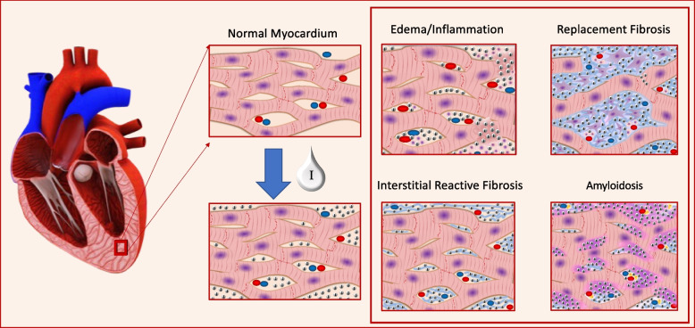
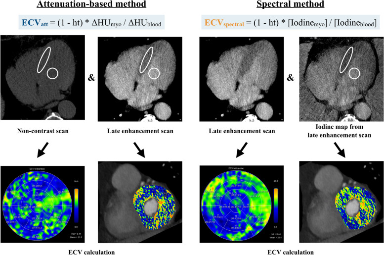
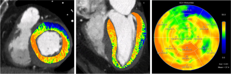
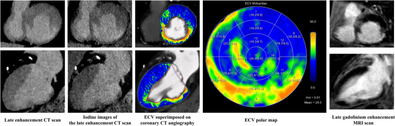
Non-invasive quantification of the extracellular volume (ECV) is a method for the evaluation of focal and diffuse myocardial fibrosis, potentially obviating the need for invasive endomyocardial biopsy. While ECV quantification with cardiac magnetic resonance imaging (ECVMRI) is already an established method, ECV quantification with CT (ECVCT) is an attractive alternative to ECVMRI, similarly using the properties of extracellular contrast media for ECV calculation. In contrast to ECVMRI, ECVCT provides a more widely available, cheaper and faster tool for ECV quantification and allows for ECV calculation also in patients with contraindications for MRI. Many studies have already shown a high correlation between ECVCT and ECVMRI and accumulating evidence suggests a prognostic value of ECVCT quantification in various cardiovascular diseases. Adding a late enhancement scan (for dual energy acquisitions) or a non-enhanced and late enhancement scan (for single-energy acquisitions) to a conventional coronary CT angiography scan improves risk stratification, requiring only minor adaptations of the contrast media and data acquisition protocols and adding only little radiation dose to the entire scan.Critical relevance statementThis article summarizes the technical principles of myocardial extracellular volume (ECV) quantification with CT, reviews the literature comparing ECVCT with ECVMRI and histopathology, and reviews the prognostic value of myocardial ECV quantification for various cardiovascular disease.Key points• Non-invasive quantification of myocardial fibrosis can be performed with CT.• Myocardial ECV quantification with CT is an alternative in patients non-eligible for MRI.• Myocardial ECV quantification with CT strongly correlates with ECV quantification using MRI.• Myocardial ECV quantification provides incremental prognostic information for various pathologies affecting the heart (e.g., cardiac amyloidosis).
Keywords: Computed tomography; Extracellular volume; Late enhancement; Myocardial fibrosis; Tissue characterization.
© 2023. European Society of Radiology (ESR).
Conflict of interest statement
H.A.: institutional grants from Bayer, Canon, Guerbet, and Siemens. Speakers bureau: Siemens.
M.E.: speakers fee from Siemens.
Figures


Similar articles
-
Myocardial Extracellular Volume Quantification by Cardiovascular Magnetic Resonance and Computed Tomography.Curr Cardiol Rep. 2018 Mar 6;20(3):15. doi: 10.1007/s11886-018-0961-3. Curr Cardiol Rep. 2018. PMID: 29511861 Free PMC article. Review.
-
Extracellular Volume Quantification With Cardiac Late Enhancement Scanning Using Dual-Source Photon-Counting Detector CT.Invest Radiol. 2022 Jun 1;57(6):406-411. doi: 10.1097/RLI.0000000000000851. Epub 2022 Jan 21. Invest Radiol. 2022. PMID: 35066531 Free PMC article.
-
Automatic quantification of the myocardial extracellular volume by cardiac computed tomography: Synthetic ECV by CCT.J Cardiovasc Comput Tomogr. 2017 May-Jun;11(3):221-226. doi: 10.1016/j.jcct.2017.02.006. Epub 2017 Feb 22. J Cardiovasc Comput Tomogr. 2017. PMID: 28268091
-
CT for the evaluation of myocardial extracellular volume with MRI as reference: a systematic review and meta-analysis.Eur Radiol. 2023 Dec;33(12):8464-8476. doi: 10.1007/s00330-023-09872-x. Epub 2023 Jun 28. Eur Radiol. 2023. PMID: 37378712
-
Myocardial extracellular volume fraction by computed tomography vs. cardiovascular magnetic resonance imaging in patients with stable chest pain.Eur Heart J Imaging Methods Pract. 2025 Mar 10;3(1):qyaf019. doi: 10.1093/ehjimp/qyaf019. eCollection 2025 Jan. Eur Heart J Imaging Methods Pract. 2025. PMID: 40066148 Free PMC article.
Cited by
-
Clinical Significance of Extracellular Volume of Myocardium (ECV) Assessed by Computed Tomography: A Systematic Review and Meta-Analysis.J Clin Med. 2025 Mar 18;14(6):2066. doi: 10.3390/jcm14062066. J Clin Med. 2025. PMID: 40142874 Free PMC article. Review.
-
In vivo 3D myocardial membrane potential mapping in humans using PET/MRI.EJNMMI Res. 2025 Jul 26;15(1):93. doi: 10.1186/s13550-025-01287-7. EJNMMI Res. 2025. PMID: 40715686 Free PMC article.
-
Quantitative assessment of early changes in myocardial extracellular volume during postoperative adjuvant chemotherapy in patients with breast cancer via dual-layer spectral detector computed tomography: a cohort study.Quant Imaging Med Surg. 2025 Jan 2;15(1):404-416. doi: 10.21037/qims-24-777. Epub 2024 Dec 30. Quant Imaging Med Surg. 2025. PMID: 39839008 Free PMC article.
-
Advanced myocardial characterization and function with cardiac CT.Int J Cardiovasc Imaging. 2024 Sep 6. doi: 10.1007/s10554-024-03229-1. Online ahead of print. Int J Cardiovasc Imaging. 2024. PMID: 39240440 Review.
-
Computer-Assisted Algorithm for Quantification of Fibrosis by Native Cardiac CT: A Pilot Study.J Clin Med. 2024 Aug 15;13(16):4807. doi: 10.3390/jcm13164807. J Clin Med. 2024. PMID: 39200950 Free PMC article.
References
Publication types
LinkOut - more resources
Full Text Sources