

A survey of Transformer applications for histopathological image analysis: New developments and future directions
- PMID: 37749595
- PMCID: PMC10518923
- DOI: 10.1186/s12938-023-01157-0
A survey of Transformer applications for histopathological image analysis: New developments and future directions
Abstract
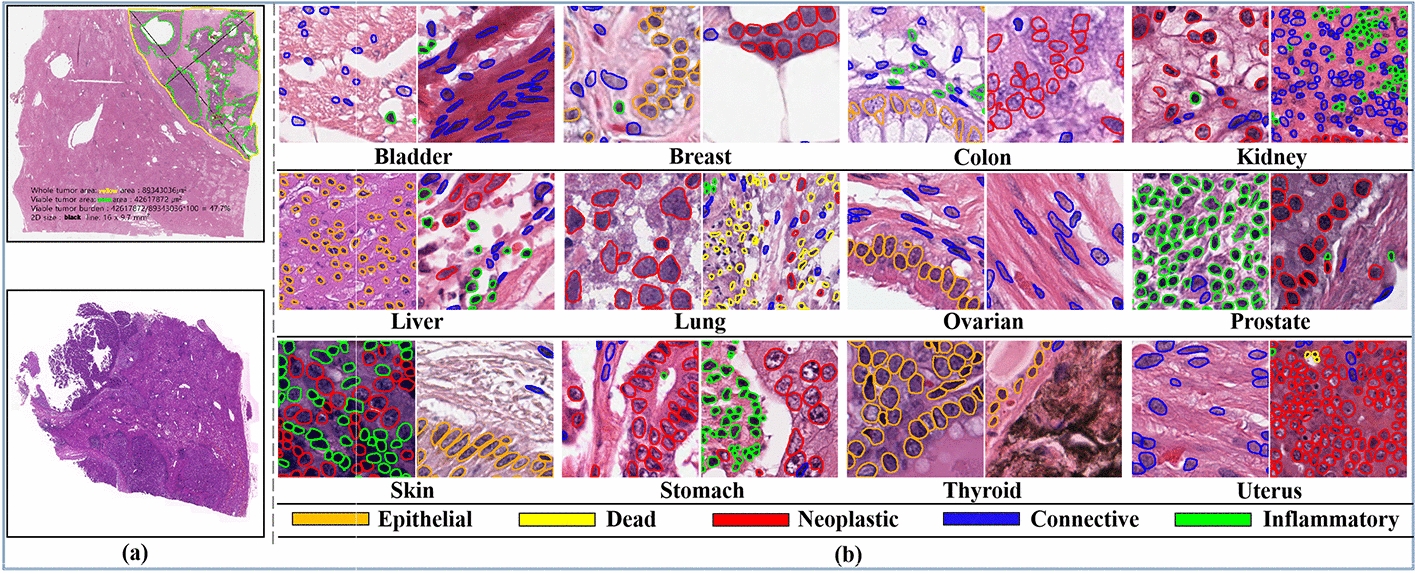
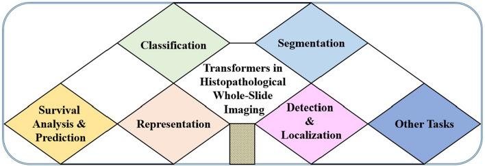
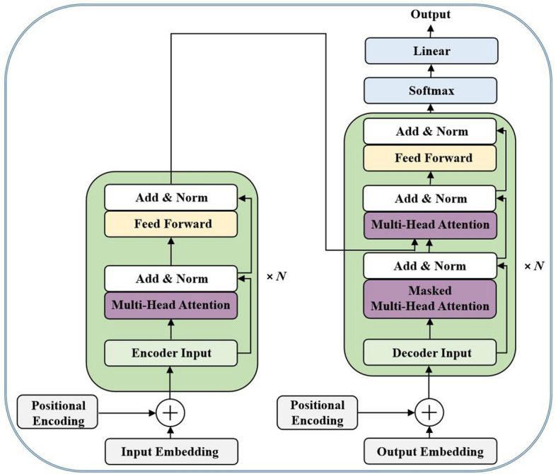
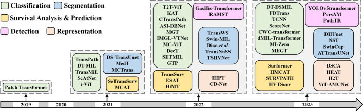
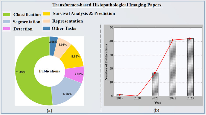
Transformers have been widely used in many computer vision challenges and have shown the capability of producing better results than convolutional neural networks (CNNs). Taking advantage of capturing long-range contextual information and learning more complex relations in the image data, Transformers have been used and applied to histopathological image processing tasks. In this survey, we make an effort to present a thorough analysis of the uses of Transformers in histopathological image analysis, covering several topics, from the newly built Transformer models to unresolved challenges. To be more precise, we first begin by outlining the fundamental principles of the attention mechanism included in Transformer models and other key frameworks. Second, we analyze Transformer-based applications in the histopathological imaging domain and provide a thorough evaluation of more than 100 research publications across different downstream tasks to cover the most recent innovations, including survival analysis and prediction, segmentation, classification, detection, and representation. Within this survey work, we also compare the performance of CNN-based techniques to Transformers based on recently published papers, highlight major challenges, and provide interesting future research directions. Despite the outstanding performance of the Transformer-based architectures in a number of papers reviewed in this survey, we anticipate that further improvements and exploration of Transformers in the histopathological imaging domain are still required in the future. We hope that this survey paper will give readers in this field of study a thorough understanding of Transformer-based techniques in histopathological image analysis, and an up-to-date paper list summary will be provided at https://github.com/S-domain/Survey-Paper .
Keywords: CNN; Digital pathology; Histopathological imaging; Survival analysis; Transformer; Whole slide image.
© 2023. BioMed Central Ltd., part of Springer Nature.
Conflict of interest statement
The authors declare that they have no competing interests.
Figures


References
-
- Shakarami A, Nicolè L, Terreran M, Dei Tos AP, Ghidoni S. Tcnn: A transformer convolutional neural network for artifact classification in whole slide images. Biomed Signal Process Control. 2023;84:104812. doi: 10.1016/j.bspc.2023.104812. - DOI
-
- Nakhli R, Moghadam PA, Mi H, Farahani H, Baras A, Gilks B, Bashashati A. Sparse multi-modal graph transformer with shared-context processing for representation learning of giga-pixel images. In: Proceedings of the IEEE/CVF Conference on Computer Vision and Pattern Recognition, pp. 11547–11557. 2023
-
- Wemmert C, Weber J, Feuerhake F, Forestier G. Deep learning for histopathological image analysis. deep learning for biomedical data analysis: techniques, approaches, and applications, 153–169. 2021.
Publication types
MeSH terms
LinkOut - more resources
Full Text Sources
Other Literature Sources
