. 2023 Sep 25;28(1):371.
doi: 10.1186/s40001-023-01340-y.

SND1 aggravates mitochondrial damage, apoptosis and extracellular matrix degradation in IL-1β-stimulated chondrocytes via PINK1/BECN1 pathway

Affiliations

SND1 aggravates mitochondrial damage, apoptosis and extracellular matrix degradation in IL-1β-stimulated chondrocytes via PINK1/BECN1 pathway

Shufeng Lin et al. Eur J Med Res. .

Abstract

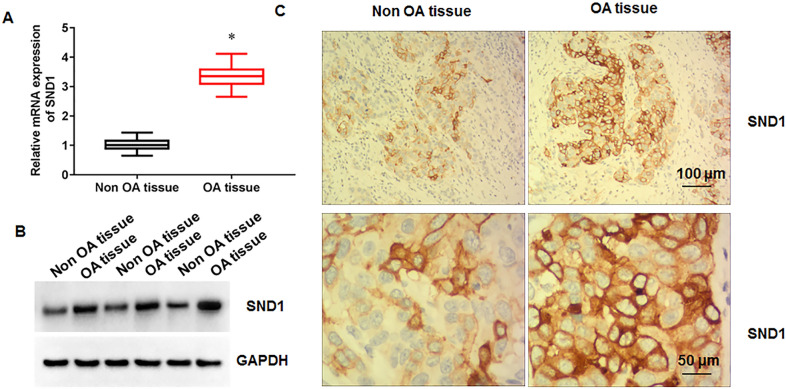
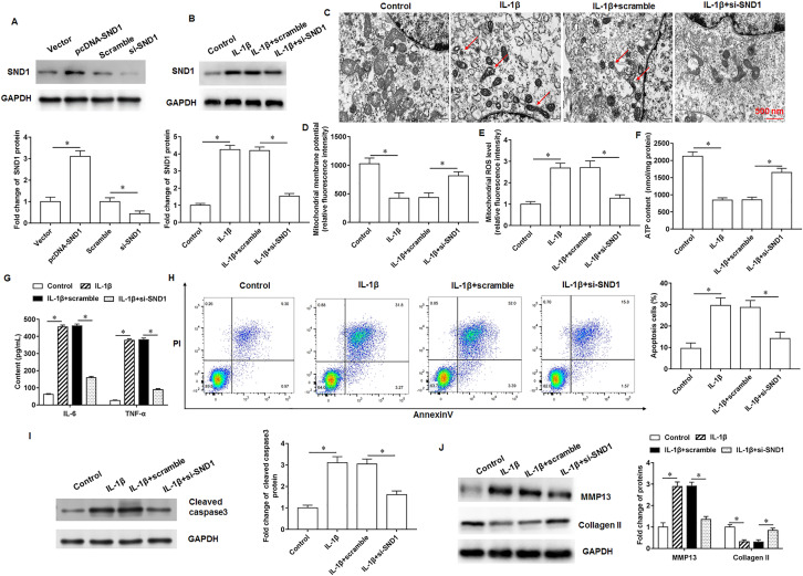
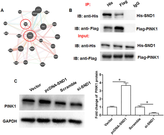
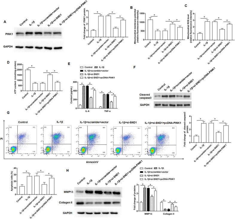
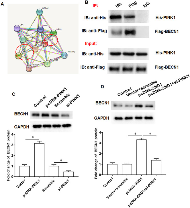
Recently, evidence has suggested a regulatory role for SND1 in osteoarthritis progression. Interestingly, we found that SND1 protein expression was increased, mitochondria were shrunken and decreased in number, mitochondrial membrane potential was decreased, mitochondrial ROS production was increased, and ATP levels were decreased in IL-1β treated mouse chondrocytes, and SND1 silencing removed these changes. Furthermore, IL-1β treatment promoted inflammatory factor secretion in chondrocytes, promoted cell apoptosis, increased MMP13 protein and inhibited collagen II protein expression, and si-SND1 inhibited the IL-1β effects. We validated the association between SND1 and PINK1 and found that PINK1 reversed the inhibitory effects of SND1 silencing on IL-1β-induced mitochondrial damage, inflammatory reaction, apoptosis and extracellular matrix degradation in mouse chondrocytes. Furthermore, we found that PINK1 upregulated BECN1 protein expression and that BECN reversed the inhibitory effects of PINK1 silencing on IL-1β-induced mitochondrial damage, inflammatory reaction, apoptosis and extracellular matrix degradation. Further mechanistic studies revealed that PINK1 inhibited the AMPK/mTOR signaling axis to aggravate IL-1β induced mouse chondrocytes injury by upregulating BECN1 protein expression. In vivo results showed that the damage to cartilage tissue was significantly alleviated in rats with osteoarthritis by knocking down SND1 expression.

Keywords: BECN1 pathway; Extracellular matrix; Mitochondrial; Osteoarthritis; SND1; The PINK1.


Conflict of interest statement

No competing interests exist in the submission of this manuscript.

Figures

Fig. 1
Fig. 1
SND1 expression in cartilage tissues of osteoarthritis patients. A. SND1 mRNA expression in knee osteoarthritis cartilage samples (N = 20) and non-osteoarthritis cartilage samples (N = 20). B. SND1 protein expression in chondrocytes isolated from knee cartilage tissues from osteoarthritis patients and fracture trauma patients. C. Immunohistochemistry was used to detect SND1 expression. N = 6. *p < 0.01
Fig. 2
Fig. 2
Effects of SND1 on IL-1β-treated chondrocytes. A. Transfection efficiency of pcDNA-SND1 or si-SND1 in chondrocytes. B. SND1 protein expression. C. Representative images of mitochondrial structure under electron microscopic field. D. Mitochondrial membrane potential levels. E. ROS content detection. F. ATP content detection. G. Inflammatory factor IL-6 and TNF-α secretion levels. H. Cell apoptosis. I, J. Cleaved caspase3, MMP13 and collagen II protein expression. N = 6. *p < 0.01
Fig. 3
Fig. 3
PINK1 is a potential binding protein for SND1. A. Online bioinformatics databases were used to predict potential binding proteins of SND1 (https://genemania.org/s; https://thebiogrid.org/). B. The binding of SND1 and PINK1 was validated by Co-IP assay. C. PINK1 protein expression. N = 6. *p < 0.01
Fig. 4
Fig. 4
SND1 affects IL-1β-treated chondrocytes through upregulation of PINK1. The IL-1β-treated chondrocytes were transfected with si-SND1 alone or together with pcDNA-PINK1. A. PINK1 protein expression. B. Mitochondrial membrane potential levels. C. ROS content detection. D. ATP content detection. E. Inflammatory factor IL-6 and TNF-α secretion levels. F. Cleaved caspase3 protein expression. G. Cell apoptosis. H. MMP13 and collagen II protein expression. N = 6. *p < 0.01
Fig. 5
Fig. 5
BECN1 is a potential binding protein forPINK1. A. Online bioinformatics databases were used to predict potential binding proteins of DRP1 (https://cn.string-db.org/). B. The binding of PINK1 and BECN1 was validated by Co-immunoprecipitation assay. C. BECN1 protein expression. D. BECN1 protein expression. N = 6. *p < 0.01
Fig. 6
Fig. 6
PINK1 affects IL-1β-treated chondrocytes through upregulation of BECN1. The IL-1β-treated chondrocytes were transfected with si-PINK1 alone or together with pcDNA-BECN1. A. BECN1 protein expression. B. AMPK and mTOR phosphorylation levels. C. Mitochondrial membrane potential levels. D. ROS content detection. E. ATP content detection. F. Inflammatory factors IL-6 and TNF-α secretion levels. G. Cell apoptosis. H, I. Cleaved caspase3, MMP13 and collagen II protein expression. N = 6. *p < 0.01
Fig. 7
Fig. 7
Effects of knockdown of SND1 expression on osteoarthritis progression in rats. Eighteen SPF male SD rats (8–10 weeks old, weighing 280–300 g) were randomly divided into three groups with 6 rats in each group, namely sham group, osteoarthritis model group and si-SND1 group. A. The SND1, PINK1 and BECN1 protein expression. B. AMPK and mTOR phosphorylation levels. C. Mitochondrial membrane potential levels. D. ROS content detection. E. The safranin O-fast green staining. F. HE staining. G. Inflammatory factors IL-6 and TNF-α secretion levels. H. Cartilage tissue score in rats. I. MMP13 and collagen II protein expression. J. Cell apoptosis. N = 6. *p < 0.01

Similar articles

Cited by

References

    1. Zhang J, et al. KLF9 and EPYC acting as feature genes for osteoarthritis and their association with immune infiltration. J Orthop Surg Res. 2022;17(1):365. doi: 10.1186/s13018-022-03247-6. - DOI - PMC - PubMed
    1. Dhlamini Q, et al. FGF1 alleviates LPS-induced acute lung injury via suppression of inflammation and oxidative stress. Mol Med. 2022;28(1):73. doi: 10.1186/s10020-022-00502-8. - DOI - PMC - PubMed
    1. Peat G, Thomas MJ. Osteoarthritis year in review 2020: epidemiology & therapy. Osteoarthritis Cartilage. 2021;29(2):180–189. doi: 10.1016/j.joca.2020.10.007. - DOI - PubMed
    1. Jacob L, Kostev K. Osteoarthritis and the incidence of fracture in the United Kingdom: a retrospective cohort study of 258,696 patients. Osteoarthritis Cartilage. 2021;29(2):215–221. doi: 10.1016/j.joca.2020.12.006. - DOI - PubMed
    1. Kan S, et al. Role of mitochondria in physiology of chondrocytes and diseases of osteoarthritis and rheumatoid arthritis. Cartilage. 2021;13:1102s–1121s. doi: 10.1177/19476035211063858. - DOI - PMC - PubMed